 Movie
Movie Controller

[English] 日本語
 Yorodumi
- PDB-6i09: Crystal structure of RlpA SPOR domain from Pseudomonas aeruginosa... -
+ Open data

- Basic information
| Entry | Database: PDB / ID: 6i09 | |||||||||
|---|---|---|---|---|---|---|---|---|---|---|
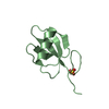
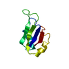
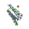
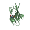
| Title | Crystal structure of RlpA SPOR domain from Pseudomonas aeruginosa in complex with denuded glycan obtained by soaking | |||||||||
 Components Components | Endolytic peptidoglycan transglycosylase RlpA | |||||||||
 Keywords Keywords | CELL CYCLE / Lytic transglycosylase / septum / SPOR domain / cell division / divisome / murein / denuded glycan | |||||||||
| Function / homology |  Function and homology information Function and homology informationLyases; Carbon-oxygen lyases; Acting on polysaccharides / lytic endotransglycosylase activity / peptidoglycan metabolic process / peptidoglycan binding / cell outer membrane / cell wall organization / plasma membrane Similarity search - Function | |||||||||
| Biological species |   Pseudomonas aeruginosa (bacteria) Pseudomonas aeruginosa (bacteria) | |||||||||
| Method |  X-RAY DIFFRACTION / SYNCHROTRON / MOLECULAR REPLACEMENT / Resolution: 1.48 Å X-RAY DIFFRACTION / SYNCHROTRON / MOLECULAR REPLACEMENT / Resolution: 1.48 Å | |||||||||
 Authors Authors | Alcorlo, M. / Hermoso, J.A. | |||||||||
| Funding support |  Spain, 1items Spain, 1items
| |||||||||
 Citation Citation | Journal: Nat Commun / Year: 2019 Title: Structural basis of denuded glycan recognition by SPOR domains in bacterial cell division. Authors: Alcorlo, M. / Dik, D.A. / De Benedetti, S. / Mahasenan, K.V. / Lee, M. / Dominguez-Gil, T. / Hesek, D. / Lastochkin, E. / Lopez, D. / Boggess, B. / Mobashery, S. / Hermoso, J.A. | |||||||||
| History |
|
- Structure visualization
| Structure viewer | Molecule: MolmilJmol/JSmol |
|---|
- Downloads & links
-Download
| PDBx/mmCIF format | 6i09.cif.gz | 55.9 KB | Display | PDBx/mmCIF format |
|---|---|---|---|---|
| PDB format | pdb6i09.ent.gz | 32.7 KB | Display | PDB format |
| PDBx/mmJSON format | 6i09.json.gz | Tree view | PDBx/mmJSON format | |
| Others |  Other downloads Other downloads |
-Validation report
| Arichive directory | https://data.pdbj.org/pub/pdb/validation_reports/i0/6i09ftp://data.pdbj.org/pub/pdb/validation_reports/i0/6i09 | HTTPS FTP |
|---|
-Related structure data
| Related structure data |  6i05SC  6i0aC  6i0nC S: Starting model for refinement C: citing same article ( |
|---|---|
| Similar structure data |

-Links
 PDBj
PDBj


- Assembly
| Deposited unit | 
| ||||||||||
|---|---|---|---|---|---|---|---|---|---|---|---|
| 1 |
| ||||||||||
| Unit cell |
| ||||||||||
| Components on special symmetry positions |
|
-Components
| #1: Protein | Mass: 8288.415 Da / Num. of mol.: 1 Source method: isolated from a genetically manipulated source Source: (gene. exp.) Pseudomonas aeruginosa (bacteria) / Gene: rlpA, PAMH19_1027 / Production host: References: UniProt: A0A0A8RDC6, UniProt: Q9X6V6*PLUS, Lyases; Carbon-oxygen lyases; Acting on polysaccharides |
|---|---|
| #2: Polysaccharide | 2-acetamido-2-deoxy-beta-D-glucopyranose-(1-4)-N-acetyl-beta-muramic acid-(1-4)-2-acetamido-2-deoxy- ...2-acetamido-2-deoxy-beta-D-glucopyranose-(1-4)-N-acetyl-beta-muramic acid-(1-4)-2-acetamido-2-deoxy-beta-D-glucopyranose-(1-4)-methyl 2-acetamido-3-O-[(1R)-1-carboxyethyl]-2-deoxy-beta-D-glucopyranoside Source method: isolated from a genetically manipulated source |
| #3: Water | ChemComp-HOH /  Mass: 18.015 Da / Num. of mol.: 73 / Source method: isolated from a natural source / Formula: H2O Mass: 18.015 Da / Num. of mol.: 73 / Source method: isolated from a natural source / Formula: H2O |
| Has ligand of interest | Y |
-Experimental details
-Experiment
| Experiment | Method: X-RAY DIFFRACTION / Number of used crystals: 1 |
|---|
- Sample preparation
| Crystal | Density Matthews: 2.72 Å3/Da / Density % sol: 54.72 % |
|---|---|
| Crystal grow | Temperature: 291 K / Method: vapor diffusion, sitting drop / Details: 0.15 M NaF and 16% (w/v) PEG3350 |
-Data collection
| Diffraction | Mean temperature: 100 K / Serial crystal experiment: N |
|---|---|
| Diffraction source | Source: SYNCHROTRON / Site: ALBA / Beamline: XALOC / Wavelength: 0.979257 Å |
| Detector | Type: DECTRIS PILATUS3 S 6M / Detector: PIXEL / Date: Oct 24, 2017 |
| Radiation | Protocol: SINGLE WAVELENGTH / Monochromatic (M) / Laue (L): M / Scattering type: x-ray |
| Radiation wavelength | Wavelength: 0.979257 Å / Relative weight: 1 |
| Reflection | Resolution: 1.48→47.934 Å / Num. obs: 15044 / % possible obs: 97.77 % / Redundancy: 7.6 % / Biso Wilson estimate: 17.78 Å2 / Rmerge(I) obs: 0.054 / Rpim(I) all: 0.02 / Net I/σ(I): 14.8 |
| Reflection shell | Resolution: 1.48→1.53 Å / Rmerge(I) obs: 0.854 / Mean I/σ(I) obs: 1.8 / Num. unique obs: 1475 / Rpim(I) all: 0.319 |
- Processing
| Software |
| ||||||||||||||||||||||||||||||||||||||||||
|---|---|---|---|---|---|---|---|---|---|---|---|---|---|---|---|---|---|---|---|---|---|---|---|---|---|---|---|---|---|---|---|---|---|---|---|---|---|---|---|---|---|---|---|
| Refinement | Method to determine structure: MOLECULAR REPLACEMENT Starting model: 6I05 Resolution: 1.48→47.93 Å / SU ML: 0.151 / Cross valid method: THROUGHOUT / σ(F): 1.34 / Phase error: 22.756 Stereochemistry target values: GeoStd + Monomer Library + CDL v1.2
| ||||||||||||||||||||||||||||||||||||||||||
| Solvent computation | Shrinkage radii: 0.9 Å / VDW probe radii: 1.11 Å / Solvent model: FLAT BULK SOLVENT MODEL | ||||||||||||||||||||||||||||||||||||||||||
| Displacement parameters | Biso mean: 29.3 Å2 | ||||||||||||||||||||||||||||||||||||||||||
| Refinement step | Cycle: LAST / Resolution: 1.48→47.93 Å
| ||||||||||||||||||||||||||||||||||||||||||
| Refine LS restraints |
| ||||||||||||||||||||||||||||||||||||||||||
| LS refinement shell |
|
