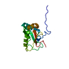
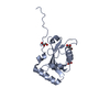
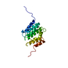
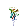
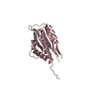
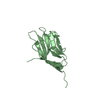
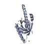
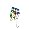
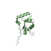
登録情報 データベース : PDB / ID : 6hylタイトル Structure of PCM1 LIR motif bound to GABARAP Pericentriolar material 1 protein,Gamma-aminobutyric acid receptor-associated protein キーワード / / / 機能・相同性 分子機能 ドメイン・相同性 構成要素
/ / / / / / / / / / / / / / / / / / / / / / / / / / / / / / / / / / / / / / / / / / / / / / / / / / / / / / / / / / / / / / / / / / / / / / / / / / / / / / / / / / / / / / / / / / / / 生物種 Homo sapiens (ヒト)手法 / / / 解像度 : 1.559 Å データ登録者 Mouilleron, S. / Wirth, M. / Zhang, W. / O'Reilly, N. / Tooze, S. / Johansen, T. / Razi, M. / Nyoni, L. / Joshi, D. ジャーナル : Nat Commun / 年 : 2019タイトル : Molecular determinants regulating selective binding of autophagy adapters and receptors to ATG8 proteins.著者 : Wirth, M. / Zhang, W. / Razi, M. / Nyoni, L. / Joshi, D. / O'Reilly, N. / Johansen, T. / Tooze, S.A. / Mouilleron, S. 履歴 登録 2018年10月22日 登録サイト / 処理サイト 改定 1.0 2019年5月8日 Provider / タイプ 改定 1.1 2019年5月15日 Group / Database referencesカテゴリ / citation_author / pdbx_database_procItem _citation.journal_volume / _citation.page_first ... _citation.journal_volume / _citation.page_first / _citation.page_last / _citation.pdbx_database_id_PubMed / _citation.title / _citation_author.identifier_ORCID / _citation_author.name 改定 1.2 2024年1月24日 Group Advisory / Data collection ... Advisory / Data collection / Database references / Refinement description カテゴリ chem_comp_atom / chem_comp_bond ... chem_comp_atom / chem_comp_bond / database_2 / pdbx_initial_refinement_model / pdbx_unobs_or_zero_occ_atoms Item / _database_2.pdbx_database_accession
すべて表示 表示を減らす
 ムービー
ムービー コントローラー


 データを開く
 基本情報
基本情報 要素
要素 キーワード
キーワード 機能・相同性情報
 Homo sapiens (ヒト)
 X線回折 /
  データ登録者
 引用
引用 構造の表示
構造の表示 ダウンロードとリンク
 その他のダウンロード


 PDBj
PDBj


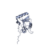
 集合体
集合体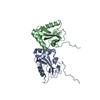

 分子量: 18.015 Da / 分子数: 234 / 由来タイプ: 天然 / 式: H2O
 試料調製
試料調製 / ビームライン: I04 / 波長: 0.9795 Å
 解析
解析