 Movie
Movie Controller


+ Open data

- Basic information
| Entry | Database: PDB / ID: 6hx3 | ||||||
|---|---|---|---|---|---|---|---|
| Title | PDX1.2/PDX1.3 complex | ||||||
 Components Components |
| ||||||
 Keywords Keywords | PLANT PROTEIN / Swinging arm lysine / Vitamin B6 / TIM-Barrel / dodecamer | ||||||
| Function / homology |  Function and homology information Function and homology informationresponse to non-ionic osmotic stress / response to lipid hydroperoxide / chlorophyll metabolic process / amine-lyase activity / pyridoxal 5'-phosphate synthase (glutamine hydrolysing) / pyridoxal 5'-phosphate synthase (glutamine hydrolysing) activity / hyperosmotic salinity response / pyridoxal 5'-phosphate biosynthetic process / pyridoxine biosynthetic process / amino acid metabolic process ...response to non-ionic osmotic stress / response to lipid hydroperoxide / chlorophyll metabolic process / amine-lyase activity / pyridoxal 5'-phosphate synthase (glutamine hydrolysing) / pyridoxal 5'-phosphate synthase (glutamine hydrolysing) activity / hyperosmotic salinity response / pyridoxal 5'-phosphate biosynthetic process / pyridoxine biosynthetic process / amino acid metabolic process / response to UV-B / response to salt stress / endomembrane system / response to oxidative stress / protein heterodimerization activity / protein homodimerization activity / plasma membrane / cytosol Similarity search - Function | ||||||
| Biological species |  | ||||||
| Method |  X-RAY DIFFRACTION / SYNCHROTRON / MOLECULAR REPLACEMENT / Resolution: 2 Å X-RAY DIFFRACTION / SYNCHROTRON / MOLECULAR REPLACEMENT / Resolution: 2 Å | ||||||
 Authors Authors | Robinson, G.C. / Kaufmann, M. / Roux, C. / Martinez-Font, J. / Hothorn, M. / Thore, S. / Fitzpatrick, T.B. | ||||||
| Funding support |  Switzerland, 1items Switzerland, 1items
| ||||||
 Citation Citation | Journal: Acta Crystallogr D Struct Biol / Year: 2019 Title: Crystal structure of the pseudoenzyme PDX1.2 in complex with its cognate enzyme PDX1.3: a total eclipse. Authors: Robinson, G.C. / Kaufmann, M. / Roux, C. / Martinez-Font, J. / Hothorn, M. / Thore, S. / Fitzpatrick, T.B. | ||||||
| History |
|
- Structure visualization
| Structure viewer | Molecule: MolmilJmol/JSmol |
|---|
- Downloads & links
-Download
| PDBx/mmCIF format | 6hx3.cif.gz | 376.8 KB | Display | PDBx/mmCIF format |
|---|---|---|---|---|
| PDB format | pdb6hx3.ent.gz | 301.9 KB | Display | PDB format |
| PDBx/mmJSON format | 6hx3.json.gz | Tree view | PDBx/mmJSON format | |
| Others |  Other downloads Other downloads |
-Validation report
| Arichive directory | https://data.pdbj.org/pub/pdb/validation_reports/hx/6hx3ftp://data.pdbj.org/pub/pdb/validation_reports/hx/6hx3 | HTTPS FTP |
|---|
-Related structure data
| Related structure data | 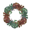 6hxgC 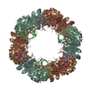 6hyeC 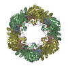 5k3vS S: Starting model for refinement C: citing same article ( |
|---|---|
| Similar structure data |


-Links
 PDBj
PDBj
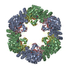
- Assembly
| Deposited unit | 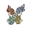
| ||||||||
|---|---|---|---|---|---|---|---|---|---|
| 1 | 
| ||||||||
| Unit cell |
|
-Components
| #1: Protein | Mass: 33874.504 Da / Num. of mol.: 4 Source method: isolated from a genetically manipulated source Source: (gene. exp.)  #2: Protein | Mass: 29071.619 Da / Num. of mol.: 4 Source method: isolated from a genetically manipulated source Source: (gene. exp.) References: UniProt: Q8L940, pyridoxal 5'-phosphate synthase (glutamine hydrolysing) #3: Chemical | ChemComp-SO4 /   Mass: 96.063 Da / Num. of mol.: 12 / Source method: obtained synthetically / Formula: SO4 Mass: 96.063 Da / Num. of mol.: 12 / Source method: obtained synthetically / Formula: SO4#4: Water | ChemComp-HOH / |  Mass: 18.015 Da / Num. of mol.: 238 / Source method: isolated from a natural source / Formula: H2O Mass: 18.015 Da / Num. of mol.: 238 / Source method: isolated from a natural source / Formula: H2O |
|---|
-Experimental details
-Experiment
| Experiment | Method: X-RAY DIFFRACTION / Number of used crystals: 1 |
|---|
- Sample preparation
| Crystal | Density Matthews: 2.58 Å3/Da / Density % sol: 52.44 % / Description: Rhombohedral |
|---|---|
| Crystal grow | Temperature: 291 K / Method: vapor diffusion, hanging drop / pH: 6.5 Details: 0.8 M Ammonium sulphate, 0.05 M MES monophydrate pH 6.5, 5 % 1,4 Dioxane 10 mM Tris pH6.8, 100 mM KCl, 3 mM DTT |
-Data collection
| Diffraction | Mean temperature: 100 K / Serial crystal experiment: N |
|---|---|
| Diffraction source | Source: SYNCHROTRON / Site: SLS / Beamline: X06SA / Wavelength: 1 Å |
| Detector | Type: DECTRIS PILATUS 2M-F / Detector: PIXEL / Date: Jul 21, 2014 |
| Radiation | Protocol: SINGLE WAVELENGTH / Monochromatic (M) / Laue (L): M / Scattering type: x-ray |
| Radiation wavelength | Wavelength: 1 Å / Relative weight: 1 |
| Reflection | Resolution: 1.84→92.33 Å / Num. obs: 118755 / % possible obs: 100 % / Redundancy: 9.6 % / Biso Wilson estimate: 26.64 Å2 / CC1/2: 0.997 / Rmerge(I) obs: 0.209 / Rpim(I) all: 0.106 / Rrim(I) all: 0.235 / Net I/σ(I): 7.7 |
| Reflection shell | Resolution: 1.84→1.87 Å / Redundancy: 9.5 % / Rmerge(I) obs: 3.698 / Mean I/σ(I) obs: 0.6 / Num. unique obs: 5897 / CC1/2: 0.129 / Rpim(I) all: 1.913 / Rrim(I) all: 4.173 / % possible all: 100 |
- Processing
| Software |
| ||||||||||||||||||||||||||||||||||||||||||||||||||||||||||||
|---|---|---|---|---|---|---|---|---|---|---|---|---|---|---|---|---|---|---|---|---|---|---|---|---|---|---|---|---|---|---|---|---|---|---|---|---|---|---|---|---|---|---|---|---|---|---|---|---|---|---|---|---|---|---|---|---|---|---|---|---|---|
| Refinement | Method to determine structure: MOLECULAR REPLACEMENT Starting model: 5K3V CHAINSAW model Resolution: 2→92.33 Å / Cor.coef. Fo:Fc: 0.948 / Cor.coef. Fo:Fc free: 0.934 / SU B: 12.916 / SU ML: 0.321 / Cross valid method: THROUGHOUT / σ(F): 0 / ESU R: 0.335 / ESU R Free: 0.21 Details: HYDROGENS HAVE BEEN ADDED IN THE RIDING POSITIONS U VALUES : REFINED INDIVIDUALLY
| ||||||||||||||||||||||||||||||||||||||||||||||||||||||||||||
| Solvent computation | Ion probe radii: 0.8 Å / Shrinkage radii: 0.8 Å / VDW probe radii: 1.2 Å | ||||||||||||||||||||||||||||||||||||||||||||||||||||||||||||
| Displacement parameters | Biso max: 100.77 Å2 / Biso mean: 32.899 Å2 / Biso min: 16.62 Å2
| ||||||||||||||||||||||||||||||||||||||||||||||||||||||||||||
| Refinement step | Cycle: final / Resolution: 2→92.33 Å
| ||||||||||||||||||||||||||||||||||||||||||||||||||||||||||||
| Refine LS restraints |
| ||||||||||||||||||||||||||||||||||||||||||||||||||||||||||||
| LS refinement shell | Resolution: 2→2.052 Å / Rfactor Rfree error: 0 / Total num. of bins used: 20
|
