[English] 日本語
Yorodumi
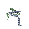
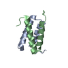
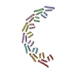
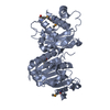
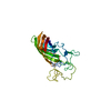
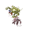
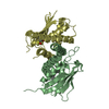
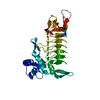
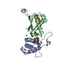
- PDB-6hs5: N-terminal domain including the conserved ImpA_N region of the Ts... -

+
Open data


ID or keywords:

Loading...

-
Basic information

Entry
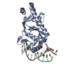
Database: PDB / ID: 6hs5
TitleN-terminal domain including the conserved ImpA_N region of the TssA component of the type VI secretion system from Burkholderia cenocepacia
ComponentsTssA
KeywordsTRANSPORT PROTEIN / alpha-helical protein / type VI secretion system component / TssA
Function / homologyType VI secretion system protein TssA-like / ImpA, N-terminal / ImpA, N-terminal, type VI secretion system / metal ion binding / Type VI secretion protein ImpA
Function and homology information
Biological speciesBurkholderia cenocepacia H111 (bacteria)
MethodX-RAY DIFFRACTION / SYNCHROTRON / SAD / Resolution: 1.8 Å
AuthorsDix, S.R. / Owen, H.J. / Sun, R. / Ahmad, A. / Shastri, S. / Spiewak, H.L. / Mosby, D.J. / Harris, M.J. / Batters, S.L. / Brooker, T.A. ...Dix, S.R. / Owen, H.J. / Sun, R. / Ahmad, A. / Shastri, S. / Spiewak, H.L. / Mosby, D.J. / Harris, M.J. / Batters, S.L. / Brooker, T.A. / Tzokov, S.B. / Sedelnikova, S.E. / Baker, P.J. / Bullough, P.A. / Rice, D.W. / Thomas, M.S.
Funding support United Kingdom, 3items
OrganizationGrant numberCountry
Biotechnology and Biological Sciences Research CouncilBB/J014443/1 United Kingdom
Biotechnology and Biological Sciences Research CouncilBB/F016840/1 United Kingdom
Higher Education Funding Council for England (HEFCE)R/151699 United Kingdom
CitationJournal: Nat Commun / Year: 2018
Title: Structural insights into the function of type VI secretion system TssA subunits.
Authors: Dix, S.R. / Owen, H.J. / Sun, R. / Ahmad, A. / Shastri, S. / Spiewak, H.L. / Mosby, D.J. / Harris, M.J. / Batters, S.L. / Brooker, T.A. / Tzokov, S.B. / Sedelnikova, S.E. / Baker, P.J. / ...Authors: Dix, S.R. / Owen, H.J. / Sun, R. / Ahmad, A. / Shastri, S. / Spiewak, H.L. / Mosby, D.J. / Harris, M.J. / Batters, S.L. / Brooker, T.A. / Tzokov, S.B. / Sedelnikova, S.E. / Baker, P.J. / Bullough, P.A. / Rice, D.W. / Thomas, M.S.
History
DepositionSep 28, 2018Deposition site: PDBE / Processing site: PDBE
Revision 1.0Nov 21, 2018Provider: repository / Type: Initial release
Revision 1.1May 15, 2024Group: Data collection / Database references / Derived calculations
Category: chem_comp_atom / chem_comp_bond ...chem_comp_atom / chem_comp_bond / database_2 / pdbx_struct_conn_angle / struct_conn
Item: _database_2.pdbx_DOI / _database_2.pdbx_database_accession ..._database_2.pdbx_DOI / _database_2.pdbx_database_accession / _pdbx_struct_conn_angle.ptnr1_auth_comp_id / _pdbx_struct_conn_angle.ptnr1_auth_seq_id / _pdbx_struct_conn_angle.ptnr1_label_asym_id / _pdbx_struct_conn_angle.ptnr1_label_atom_id / _pdbx_struct_conn_angle.ptnr1_label_comp_id / _pdbx_struct_conn_angle.ptnr1_label_seq_id / _pdbx_struct_conn_angle.ptnr1_symmetry / _pdbx_struct_conn_angle.ptnr2_symmetry / _pdbx_struct_conn_angle.ptnr3_auth_comp_id / _pdbx_struct_conn_angle.ptnr3_auth_seq_id / _pdbx_struct_conn_angle.ptnr3_label_asym_id / _pdbx_struct_conn_angle.ptnr3_label_atom_id / _pdbx_struct_conn_angle.ptnr3_label_comp_id / _pdbx_struct_conn_angle.ptnr3_label_seq_id / _pdbx_struct_conn_angle.ptnr3_symmetry / _pdbx_struct_conn_angle.value / _struct_conn.pdbx_dist_value / _struct_conn.ptnr1_auth_comp_id / _struct_conn.ptnr1_auth_seq_id / _struct_conn.ptnr1_label_asym_id / _struct_conn.ptnr1_label_atom_id / _struct_conn.ptnr1_label_comp_id / _struct_conn.ptnr1_label_seq_id / _struct_conn.ptnr2_auth_comp_id / _struct_conn.ptnr2_auth_seq_id / _struct_conn.ptnr2_label_asym_id / _struct_conn.ptnr2_label_atom_id / _struct_conn.ptnr2_label_comp_id / _struct_conn.ptnr2_symmetry

-
Structure visualization

Structure viewerMolecule:
MolmilJmol/JSmol

Downloads & links

-
Assembly

Deposited unit
A: TssA
hetero molecules


Theoretical massNumber of molelcules
Total (without water)30,9574
Polymers30,8151
Non-polymers1423
Water2,144119
1


  • Idetical with deposited unit
  • defined by author&software
  • Evidence: gel filtration
TypeNameSymmetry operationNumber
identity operation1_555x,y,z1
Buried area250 Å2
ΔGint-8 kcal/mol
Surface area13460 Å2
MethodPISA
Unit cell
Length a, b, c (Å)49.670, 125.740, 45.560
Angle α, β, γ (deg.)90.00, 90.00, 90.00
Int Tables number18
Space group name H-MP21212
Components on special symmetry positions
IDModelComponents
11A-301-

CA

-
Components

#1: Protein TssA


Mass: 30815.059 Da / Num. of mol.: 1
Source method: isolated from a genetically manipulated source
Details: N-terminal His-tag followed by a thrombin cleavage site - MGSSHHHHHHSSGLVPRGSH Construct comprises residues 1-255 of full-length protein (total 373 residues)
Source: (gene. exp.) Burkholderia cenocepacia H111 (bacteria)
Gene: I35_RS01755 / Plasmid: pET14b / Production host: Escherichia coli BL21(DE3) (bacteria) / References: UniProt: A0A1V2W6E8
#2: Chemical ChemComp-CA / CALCIUM ION


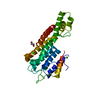
Mass: 40.078 Da / Num. of mol.: 2 / Source method: obtained synthetically / Formula: Ca
#3: Chemical ChemComp-EDO / 1,2-ETHANEDIOL / ETHYLENE GLYCOL


Mass: 62.068 Da / Num. of mol.: 1 / Source method: obtained synthetically / Formula: C2H6O2
#4: Water ChemComp-HOH / water


Mass: 18.015 Da / Num. of mol.: 119 / Source method: isolated from a natural source / Formula: H2O

-
Experimental details

-
Experiment

ExperimentMethod: X-RAY DIFFRACTION / Number of used crystals: 1

-
Sample preparation

CrystalDensity Matthews: 2.31 Å3/Da / Density % sol: 46.72 %
Crystal growTemperature: 290 K / Method: vapor diffusion, sitting drop
Details: 0.16M calcium acetate, 0.08M sodium cacodylate pH6.5, 14.4% (w/v) PEG8000, 20% (v/v) glycerol

-
Data collection

DiffractionMean temperature: 100 K
Diffraction sourceSource: SYNCHROTRON / Site: Diamond / Beamline: I02 / Wavelength: 1.04434 Å
DetectorType: DECTRIS PILATUS 6M-F / Detector: PIXEL / Date: Jul 24, 2013
RadiationProtocol: SINGLE WAVELENGTH / Monochromatic (M) / Laue (L): M / Scattering type: x-ray
Radiation wavelengthWavelength: 1.04434 Å / Relative weight: 1
ReflectionResolution: 1.8→49.67 Å / Num. obs: 27244 / % possible obs: 99.8 % / Redundancy: 5.4 % / CC1/2: 0.999 / Rmerge(I) obs: 0.043 / Rpim(I) all: 0.022 / Net I/σ(I): 21.2
Reflection shellResolution: 1.8→1.85 Å / Redundancy: 5.3 % / Rmerge(I) obs: 0.344 / Mean I/σ(I) obs: 4.4 / Num. unique obs: 1966 / CC1/2: 0.909 / Rpim(I) all: 0.177 / % possible all: 99

-
Processing

Software
NameVersionClassification
REFMAC5.8.0135refinement
xia2data reduction
xia2data scaling
SHELXCDphasing
RefinementMethod to determine structure: SAD / Resolution: 1.8→46.21 Å / Cor.coef. Fo:Fc: 0.96 / Cor.coef. Fo:Fc free: 0.939 / SU B: 2.567 / SU ML: 0.079 / Cross valid method: THROUGHOUT / ESU R: 0.113 / ESU R Free: 0.119 / Details: HYDROGENS HAVE BEEN ADDED IN THE RIDING POSITIONS
RfactorNum. reflection% reflectionSelection details
Rfree0.22594 1381 5.1 %RANDOM
Rwork0.17719 ---
obs0.17966 25817 99.74 %-
Solvent computationIon probe radii: 0.8 Å / Shrinkage radii: 0.8 Å / VDW probe radii: 1.2 Å
Displacement parametersBiso mean: 29.614 Å2
Baniso -1Baniso -2Baniso -3
1-0.59 Å20 Å20 Å2
2---0.33 Å20 Å2
3----0.26 Å2
Refinement stepCycle: 1 / Resolution: 1.8→46.21 Å
ProteinNucleic acidLigandSolventTotal
Num. atoms1971 0 6 119 2096
Refine LS restraints
Refine-IDTypeDev idealDev ideal targetNumber
X-RAY DIFFRACTIONr_bond_refined_d0.0160.0192041
X-RAY DIFFRACTIONr_bond_other_d0.0020.021890
X-RAY DIFFRACTIONr_angle_refined_deg1.5671.9532774
X-RAY DIFFRACTIONr_angle_other_deg1.00734337
X-RAY DIFFRACTIONr_dihedral_angle_1_deg5.3235253
X-RAY DIFFRACTIONr_dihedral_angle_2_deg35.3223.394109
X-RAY DIFFRACTIONr_dihedral_angle_3_deg13.09115324
X-RAY DIFFRACTIONr_dihedral_angle_4_deg18.6391521
X-RAY DIFFRACTIONr_chiral_restr0.1030.2302
X-RAY DIFFRACTIONr_gen_planes_refined0.0080.0212343
X-RAY DIFFRACTIONr_gen_planes_other0.0010.02490
X-RAY DIFFRACTIONr_nbd_refined
X-RAY DIFFRACTIONr_nbd_other
X-RAY DIFFRACTIONr_nbtor_refined
X-RAY DIFFRACTIONr_nbtor_other
X-RAY DIFFRACTIONr_xyhbond_nbd_refined
X-RAY DIFFRACTIONr_xyhbond_nbd_other
X-RAY DIFFRACTIONr_metal_ion_refined
X-RAY DIFFRACTIONr_metal_ion_other
X-RAY DIFFRACTIONr_symmetry_vdw_refined
X-RAY DIFFRACTIONr_symmetry_vdw_other
X-RAY DIFFRACTIONr_symmetry_hbond_refined
X-RAY DIFFRACTIONr_symmetry_hbond_other
X-RAY DIFFRACTIONr_symmetry_metal_ion_refined
X-RAY DIFFRACTIONr_symmetry_metal_ion_other
X-RAY DIFFRACTIONr_mcbond_it34.9912.3611000
X-RAY DIFFRACTIONr_mcbond_other35.0012.357999
X-RAY DIFFRACTIONr_mcangle_it25.9023.4171248
X-RAY DIFFRACTIONr_mcangle_other25.8933.4181249
X-RAY DIFFRACTIONr_scbond_it53.9013.4561041
X-RAY DIFFRACTIONr_scbond_other53.8993.4571041
X-RAY DIFFRACTIONr_scangle_it
X-RAY DIFFRACTIONr_scangle_other36.6984.471524
X-RAY DIFFRACTIONr_long_range_B_refined38.50920.6642491
X-RAY DIFFRACTIONr_long_range_B_other38.95720.4882432
X-RAY DIFFRACTIONr_rigid_bond_restr
X-RAY DIFFRACTIONr_sphericity_free
X-RAY DIFFRACTIONr_sphericity_bonded
LS refinement shellResolution: 1.8→1.847 Å / Total num. of bins used: 20
RfactorNum. reflection% reflection
Rfree0.282 96 -
Rwork0.235 1861 -
obs--98.84 %

+
About Yorodumi

-
News

-
Feb 9, 2022. New format data for meta-information of EMDB entries

New format data for meta-information of EMDB entries

  • Version 3 of the EMDB header file is now the official format.
  • The previous official version 1.9 will be removed from the archive.

Related info.:EMDB header

External links:wwPDB to switch to version 3 of the EMDB data model

-
Aug 12, 2020. Covid-19 info

Covid-19 info

URL: https://pdbjlvh1.pdbj.org/emnavi/covid19.php

New page: Covid-19 featured information page in EM Navigator.

Related info.:Covid-19 info / Mar 5, 2020. Novel coronavirus structure data

+
Mar 5, 2020. Novel coronavirus structure data

Novel coronavirus structure data

Related info.:Yorodumi Speices / Aug 12, 2020. Covid-19 info

External links:COVID-19 featured content - PDBj / Molecule of the Month (242):Coronavirus Proteases

+
Jan 31, 2019. EMDB accession codes are about to change! (news from PDBe EMDB page)

EMDB accession codes are about to change! (news from PDBe EMDB page)

  • The allocation of 4 digits for EMDB accession codes will soon come to an end. Whilst these codes will remain in use, new EMDB accession codes will include an additional digit and will expand incrementally as the available range of codes is exhausted. The current 4-digit format prefixed with “EMD-” (i.e. EMD-XXXX) will advance to a 5-digit format (i.e. EMD-XXXXX), and so on. It is currently estimated that the 4-digit codes will be depleted around Spring 2019, at which point the 5-digit format will come into force.
  • The EM Navigator/Yorodumi systems omit the EMD- prefix.

Related info.:Q: What is EMD? / ID/Accession-code notation in Yorodumi/EM Navigator

External links:EMDB Accession Codes are Changing Soon! / Contact to PDBj

+
Jul 12, 2017. Major update of PDB

Major update of PDB

  • wwPDB released updated PDB data conforming to the new PDBx/mmCIF dictionary.
  • This is a major update changing the version number from 4 to 5, and with Remediation, in which all the entries are updated.
  • In this update, many items about electron microscopy experimental information are reorganized (e.g. em_software).
  • Now, EM Navigator and Yorodumi are based on the updated data.

External links:wwPDB Remediation / Enriched Model Files Conforming to OneDep Data Standards Now Available in the PDB FTP Archive

-
Yorodumi

Thousand views of thousand structures

  • Yorodumi is a browser for structure data from EMDB, PDB, SASBDB, etc.
  • This page is also the successor to EM Navigator detail page, and also detail information page/front-end page for Omokage search.
  • The word "yorodu" (or yorozu) is an old Japanese word meaning "ten thousand". "mi" (miru) is to see.

Related info.:EMDB / PDB / SASBDB / Comparison of 3 databanks / Yorodumi Search / Aug 31, 2016. New EM Navigator & Yorodumi / Yorodumi Papers / Jmol/JSmol / Function and homology information / Changes in new EM Navigator and Yorodumi

Read more