 Movie
Movie Controller

[English] 日本語
 Yorodumi
- PDB-6hl7: Crystal structure of truncated aspartate transcarbamoylase from P... -
+ Open data

- Basic information
| Entry | Database: PDB / ID: 6hl7 | |||||||||
|---|---|---|---|---|---|---|---|---|---|---|
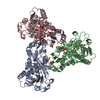
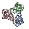
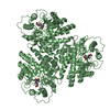
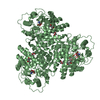
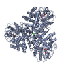
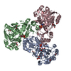
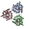
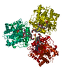
| Title | Crystal structure of truncated aspartate transcarbamoylase from Plasmodium falciparum with mutated active site (R109A/K138A) and N-carbamoyl-L-phosphate bound | |||||||||
 Components Components | Aspartate transcarbamoylase | |||||||||
 Keywords Keywords | TRANSFERASE / Falciparum Malaria Pyrimidine biosynthesis Trimer Mutant TRANSFERASE carbamoyl-phospahte | |||||||||
| Function / homology |  Function and homology information Function and homology informationaspartate carbamoyltransferase / aspartate carbamoyltransferase activity / amino acid metabolic process / amino acid binding / 'de novo' UMP biosynthetic process / 'de novo' pyrimidine nucleobase biosynthetic process Similarity search - Function | |||||||||
| Biological species |  | |||||||||
| Method |  X-RAY DIFFRACTION / SYNCHROTRON / MOLECULAR REPLACEMENT / Resolution: 2.5 Å X-RAY DIFFRACTION / SYNCHROTRON / MOLECULAR REPLACEMENT / Resolution: 2.5 Å | |||||||||
 Authors Authors | Bosch, S.S. / Lunev, S. / Wrenger, C. / Groves, M.R. | |||||||||
| Funding support |  Brazil, 2items Brazil, 2items
| |||||||||
 Citation Citation | Journal: Acs Infect Dis. / Year: 2020 Title: Molecular Target Validation of Aspartate Transcarbamoylase fromPlasmodium falciparumby Torin 2. Authors: Bosch, S.S. / Lunev, S. / Batista, F.A. / Linzke, M. / Kronenberger, T. / Domling, A.S.S. / Groves, M.R. / Wrenger, C. #1: Journal: Acta Crystallogr F Struct Biol Commun / Year: 2016Title: Crystal structure of truncated aspartate transcarbamoylase from Plasmodium falciparum. Authors: Lunev, S. / Bosch, S.S. / de Assis Batista, F. / Wrenger, C. / Groves, M.R. | |||||||||
| History |
|
- Structure visualization
| Structure viewer | Molecule: MolmilJmol/JSmol |
|---|
- Downloads & links
-Download
| PDBx/mmCIF format | 6hl7.cif.gz | 205.7 KB | Display | PDBx/mmCIF format |
|---|---|---|---|---|
| PDB format | pdb6hl7.ent.gz | 165 KB | Display | PDB format |
| PDBx/mmJSON format | 6hl7.json.gz | Tree view | PDBx/mmJSON format | |
| Others |  Other downloads Other downloads |
-Validation report
| Arichive directory | https://data.pdbj.org/pub/pdb/validation_reports/hl/6hl7ftp://data.pdbj.org/pub/pdb/validation_reports/hl/6hl7 | HTTPS FTP |
|---|
-Related structure data
| Related structure data |  5ilqS S: Starting model for refinement |
|---|---|
| Similar structure data |
-Links
 PDBj
PDBj
- Assembly
| Deposited unit | 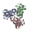
| ||||||||
|---|---|---|---|---|---|---|---|---|---|
| 1 |
| ||||||||
| Unit cell |
|
-Components
| #1: Protein | Mass: 40119.465 Da / Num. of mol.: 3 / Mutation: R109A, K138A Source method: isolated from a genetically manipulated source Source: (gene. exp.) Gene: ATCase / Production host:  #2: Chemical |   Mass: 141.020 Da / Num. of mol.: 2 / Source method: obtained synthetically / Formula: CH4NO5P Mass: 141.020 Da / Num. of mol.: 2 / Source method: obtained synthetically / Formula: CH4NO5P#3: Water | ChemComp-HOH / |  Mass: 18.015 Da / Num. of mol.: 42 / Source method: isolated from a natural source / Formula: H2O Mass: 18.015 Da / Num. of mol.: 42 / Source method: isolated from a natural source / Formula: H2O |
|---|
-Experimental details
-Experiment
| Experiment | Method: X-RAY DIFFRACTION / Number of used crystals: 1 |
|---|
- Sample preparation
| Crystal | Density Matthews: 3.06 Å3/Da / Density % sol: 59.76 % |
|---|---|
| Crystal grow | Temperature: 293 K / Method: vapor diffusion Details: 0.2M Potassium citrate tribasic monohydrate 20% w/v PEG 3350 |
-Data collection
| Diffraction | Mean temperature: 100 K |
|---|---|
| Diffraction source | Source: SYNCHROTRON / Site: ESRF  / Beamline: ID23-1 / Wavelength: 0.976 Å / Beamline: ID23-1 / Wavelength: 0.976 Å |
| Detector | Type: DECTRIS PILATUS 6M / Detector: PIXEL / Date: Apr 3, 2016 |
| Radiation | Protocol: SINGLE WAVELENGTH / Monochromatic (M) / Laue (L): M / Scattering type: x-ray |
| Radiation wavelength | Wavelength: 0.976 Å / Relative weight: 1 |
| Reflection | Resolution: 2.5→57.61 Å / Num. obs: 46404 / % possible obs: 97.6 % / Observed criterion σ(I): 1.3 / Redundancy: 3.16 % / CC1/2: 0.999 / Rrim(I) all: 0.066 / Net I/σ(I): 12.95 |
| Reflection shell | Resolution: 2.5→2.64 Å / Redundancy: 3.21 % / Num. unique obs: 7154 / CC1/2: 0.619 / Rrim(I) all: 0.957 / % possible all: 94 |
- Processing
| Software |
| |||||||||||||||||||||||||||||||||||||||||||||||||||||||||||||||||||||||||||
|---|---|---|---|---|---|---|---|---|---|---|---|---|---|---|---|---|---|---|---|---|---|---|---|---|---|---|---|---|---|---|---|---|---|---|---|---|---|---|---|---|---|---|---|---|---|---|---|---|---|---|---|---|---|---|---|---|---|---|---|---|---|---|---|---|---|---|---|---|---|---|---|---|---|---|---|---|
| Refinement | Method to determine structure: MOLECULAR REPLACEMENT Starting model: 5ilq Resolution: 2.5→57.61 Å / Cor.coef. Fo:Fc: 0.956 / Cor.coef. Fo:Fc free: 0.928 / Cross valid method: THROUGHOUT / σ(F): 0 / ESU R: 0.432 / ESU R Free: 0.283 Details: HYDROGENS HAVE BEEN ADDED IN THE RIDING POSITIONS U VALUES : REFINED INDIVIDUALLY
| |||||||||||||||||||||||||||||||||||||||||||||||||||||||||||||||||||||||||||
| Solvent computation | Ion probe radii: 0.8 Å / Shrinkage radii: 0.8 Å / VDW probe radii: 1.2 Å | |||||||||||||||||||||||||||||||||||||||||||||||||||||||||||||||||||||||||||
| Displacement parameters | Biso max: 163.5 Å2 / Biso mean: 65.452 Å2 / Biso min: 28.67 Å2
| |||||||||||||||||||||||||||||||||||||||||||||||||||||||||||||||||||||||||||
| Refinement step | Cycle: final / Resolution: 2.5→57.61 Å
| |||||||||||||||||||||||||||||||||||||||||||||||||||||||||||||||||||||||||||
| Refine LS restraints |
| |||||||||||||||||||||||||||||||||||||||||||||||||||||||||||||||||||||||||||
| LS refinement shell | Resolution: 2.502→2.567 Å / Total num. of bins used: 20
|
