 Movie
Movie Controller

[English] 日本語
 Yorodumi
- PDB-5iln: Crystal structure of Aspartate Transcarbamoylase from Plasmodium ... -
+ Open data

- Basic information
| Entry | Database: PDB / ID: 5iln | ||||||
|---|---|---|---|---|---|---|---|
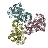
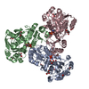
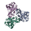
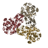
| Title | Crystal structure of Aspartate Transcarbamoylase from Plasmodium falciparum (PfATC) with bound citrate | ||||||
 Components Components | Aspartate carbamoyltransferase | ||||||
 Keywords Keywords | TRANSFERASE / Plasmodium / Malaria / Aspartate / Pyrimidine Biosynthesis | ||||||
| Function / homology |  Function and homology information Function and homology information: / aspartate carbamoyltransferase / aspartate carbamoyltransferase activity / amino acid metabolic process / amino acid binding / membrane => GO:0016020 / 'de novo' pyrimidine nucleobase biosynthetic process / cytoplasm / cytosol Similarity search - Function | ||||||
| Biological species |  | ||||||
| Method |  X-RAY DIFFRACTION / SYNCHROTRON / MOLECULAR REPLACEMENT / Resolution: 2.21 Å X-RAY DIFFRACTION / SYNCHROTRON / MOLECULAR REPLACEMENT / Resolution: 2.21 Å | ||||||
 Authors Authors | Lunev, S. / Bosch, S.S. / Batista, F.D.A. / Wrenger, C. / Groves, M.R. | ||||||
 Citation Citation | Journal: To be published Title: Crystal structure of truncated Aspartate Transcarbamoylase from Plasmodium falciparum Authors: Lunev, S. / Bosch, S.S. / Batista, F.D.A. / Wrenger, C. / Groves, M.R. | ||||||
| History |
|
- Structure visualization
| Structure viewer | Molecule: MolmilJmol/JSmol |
|---|
- Downloads & links
-Download
| PDBx/mmCIF format | 5iln.cif.gz | 314.1 KB | Display | PDBx/mmCIF format |
|---|---|---|---|---|
| PDB format | pdb5iln.ent.gz | 245.5 KB | Display | PDB format |
| PDBx/mmJSON format | 5iln.json.gz | Tree view | PDBx/mmJSON format | |
| Others |  Other downloads Other downloads |
-Validation report
| Arichive directory | https://data.pdbj.org/pub/pdb/validation_reports/il/5ilnftp://data.pdbj.org/pub/pdb/validation_reports/il/5iln | HTTPS FTP |
|---|
-Related structure data
| Similar structure data |
|---|

-Links
 PDBj
PDBj


- Assembly
| Deposited unit | 
| |||||||||||||||||||||||||||||||||||||||||||||||||||||||||||||||||||||||||||||||||||||||||||||||||||||||||||||||||||||||||||||||||||||||||||||||||||||||||||||||||||||||||||||||||||||||||||||||||||||||||||||||||||||||||||||||||
|---|---|---|---|---|---|---|---|---|---|---|---|---|---|---|---|---|---|---|---|---|---|---|---|---|---|---|---|---|---|---|---|---|---|---|---|---|---|---|---|---|---|---|---|---|---|---|---|---|---|---|---|---|---|---|---|---|---|---|---|---|---|---|---|---|---|---|---|---|---|---|---|---|---|---|---|---|---|---|---|---|---|---|---|---|---|---|---|---|---|---|---|---|---|---|---|---|---|---|---|---|---|---|---|---|---|---|---|---|---|---|---|---|---|---|---|---|---|---|---|---|---|---|---|---|---|---|---|---|---|---|---|---|---|---|---|---|---|---|---|---|---|---|---|---|---|---|---|---|---|---|---|---|---|---|---|---|---|---|---|---|---|---|---|---|---|---|---|---|---|---|---|---|---|---|---|---|---|---|---|---|---|---|---|---|---|---|---|---|---|---|---|---|---|---|---|---|---|---|---|---|---|---|---|---|---|---|---|---|---|---|---|---|---|---|---|---|---|---|---|---|---|---|---|---|---|---|
| 1 |
| |||||||||||||||||||||||||||||||||||||||||||||||||||||||||||||||||||||||||||||||||||||||||||||||||||||||||||||||||||||||||||||||||||||||||||||||||||||||||||||||||||||||||||||||||||||||||||||||||||||||||||||||||||||||||||||||||
| Unit cell |
| |||||||||||||||||||||||||||||||||||||||||||||||||||||||||||||||||||||||||||||||||||||||||||||||||||||||||||||||||||||||||||||||||||||||||||||||||||||||||||||||||||||||||||||||||||||||||||||||||||||||||||||||||||||||||||||||||
| Noncrystallographic symmetry (NCS) | NCS domain:
NCS domain segments:
NCS ensembles :
NCS oper:
|
-Components
| #1: Protein | Mass: 44539.012 Da / Num. of mol.: 3 Source method: isolated from a genetically manipulated source Source: (gene. exp.) Gene: atcasE, MAL13P1.221 / Plasmid: pASK-IBA3-PfATC-full / Details (production host): full length pfATC / Cell line (production host): Rosetta 2(DE3) pLysS / Production host:  #2: Chemical | ChemComp-GOL /   Mass: 92.094 Da / Num. of mol.: 4 / Source method: obtained synthetically / Formula: C3H8O3 Mass: 92.094 Da / Num. of mol.: 4 / Source method: obtained synthetically / Formula: C3H8O3#3: Chemical |   Mass: 94.971 Da / Num. of mol.: 3 / Source method: obtained synthetically / Formula: PO4 Mass: 94.971 Da / Num. of mol.: 3 / Source method: obtained synthetically / Formula: PO4#4: Chemical | ChemComp-FLC / |   Mass: 189.100 Da / Num. of mol.: 1 / Source method: obtained synthetically / Formula: C6H5O7 Mass: 189.100 Da / Num. of mol.: 1 / Source method: obtained synthetically / Formula: C6H5O7#5: Water | ChemComp-HOH / |  Mass: 18.015 Da / Num. of mol.: 166 / Source method: isolated from a natural source / Formula: H2O Mass: 18.015 Da / Num. of mol.: 166 / Source method: isolated from a natural source / Formula: H2OHas protein modification | Y | |
|---|
-Experimental details
-Experiment
| Experiment | Method: X-RAY DIFFRACTION / Number of used crystals: 1 |
|---|
- Sample preparation
| Crystal | Density Matthews: 2.32 Å3/Da / Density % sol: 47 % |
|---|---|
| Crystal grow | Temperature: 293 K / Method: vapor diffusion, sitting drop Details: 0.2 M Potassium citrate tribasic monohydrate, 20 % w/v PEG 3350 |
-Data collection
| Diffraction | Mean temperature: 100 K / Ambient temp details: Oxford Cryostream - 700 series |
|---|---|
| Diffraction source | Source: SYNCHROTRON / Site: ESRF  / Beamline: MASSIF-1 / Wavelength: 0.965 Å / Beamline: MASSIF-1 / Wavelength: 0.965 Å |
| Detector | Type: DECTRIS PILATUS 2M / Detector: PIXEL / Date: Feb 2, 2015 |
| Radiation | Monochromator: Diamond / Protocol: SINGLE WAVELENGTH / Monochromatic (M) / Laue (L): M / Scattering type: x-ray |
| Radiation wavelength | Wavelength: 0.965 Å / Relative weight: 1 |
| Reflection | Resolution: 2.21→45.1 Å / Num. obs: 69115 / % possible obs: 97.7 % / Redundancy: 2.9 % / CC1/2: 0.984 / Rmerge(I) obs: 0.108 / Rsym value: 0.112 / Net I/σ(I): 5 |
| Reflection shell | Resolution: 2.21→2.29 Å / Redundancy: 2.2 % / Rmerge(I) obs: 0.8 / Mean I/σ(I) obs: 0.8 / % possible all: 83.8 |
- Processing
| Software |
| ||||||||||||||||||||||||||||||||||||||||||||||||||||||||||||||||||||||||||||||||||||||||||||||||||||||||||||||||||||||||||||||||||||||||||||||||||||||||||||||||||||||||||||||||||||||
|---|---|---|---|---|---|---|---|---|---|---|---|---|---|---|---|---|---|---|---|---|---|---|---|---|---|---|---|---|---|---|---|---|---|---|---|---|---|---|---|---|---|---|---|---|---|---|---|---|---|---|---|---|---|---|---|---|---|---|---|---|---|---|---|---|---|---|---|---|---|---|---|---|---|---|---|---|---|---|---|---|---|---|---|---|---|---|---|---|---|---|---|---|---|---|---|---|---|---|---|---|---|---|---|---|---|---|---|---|---|---|---|---|---|---|---|---|---|---|---|---|---|---|---|---|---|---|---|---|---|---|---|---|---|---|---|---|---|---|---|---|---|---|---|---|---|---|---|---|---|---|---|---|---|---|---|---|---|---|---|---|---|---|---|---|---|---|---|---|---|---|---|---|---|---|---|---|---|---|---|---|---|---|---|
| Refinement | Method to determine structure: MOLECULAR REPLACEMENT Starting model: PfATC-Met3 Resolution: 2.21→45.1 Å / Cor.coef. Fo:Fc: 0.967 / Cor.coef. Fo:Fc free: 0.943 / SU B: 13.546 / SU ML: 0.18 / Cross valid method: FREE R-VALUE / ESU R: 0.209 / ESU R Free: 0.19 / Details: HYDROGENS HAVE BEEN ADDED IN THE RIDING POSITIONS
| ||||||||||||||||||||||||||||||||||||||||||||||||||||||||||||||||||||||||||||||||||||||||||||||||||||||||||||||||||||||||||||||||||||||||||||||||||||||||||||||||||||||||||||||||||||||
| Solvent computation | Ion probe radii: 0.8 Å / Shrinkage radii: 0.8 Å / VDW probe radii: 1.2 Å | ||||||||||||||||||||||||||||||||||||||||||||||||||||||||||||||||||||||||||||||||||||||||||||||||||||||||||||||||||||||||||||||||||||||||||||||||||||||||||||||||||||||||||||||||||||||
| Displacement parameters | Biso mean: 51.616 Å2
| ||||||||||||||||||||||||||||||||||||||||||||||||||||||||||||||||||||||||||||||||||||||||||||||||||||||||||||||||||||||||||||||||||||||||||||||||||||||||||||||||||||||||||||||||||||||
| Refinement step | Cycle: 1 / Resolution: 2.21→45.1 Å
| ||||||||||||||||||||||||||||||||||||||||||||||||||||||||||||||||||||||||||||||||||||||||||||||||||||||||||||||||||||||||||||||||||||||||||||||||||||||||||||||||||||||||||||||||||||||
| Refine LS restraints |
|