 Movie
Movie Controller

[English] 日本語
 Yorodumi
- PDB-4wvc: Crystal structure of GH63 mannosylglycerate hydrolase from Thermu... -
+ Open data

- Basic information
| Entry | Database: PDB / ID: 4wvc | ||||||
|---|---|---|---|---|---|---|---|
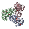
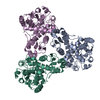
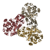
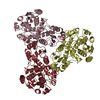
| Title | Crystal structure of GH63 mannosylglycerate hydrolase from Thermus thermophilus HB8 in complex with Tris and D-glycerate | ||||||
 Components Components | Uncharacterized protein | ||||||
 Keywords Keywords | HYDROLASE / GH63 / (alpha/alpha)6-barrel | ||||||
| Function / homology |  Function and homology information Function and homology informationGlc3Man9GlcNAc2 oligosaccharide glucosidase activity / oligosaccharide metabolic process / protein N-linked glycosylation Similarity search - Function | ||||||
| Biological species |   Thermus thermophilus HB8 (bacteria) Thermus thermophilus HB8 (bacteria) | ||||||
| Method |  X-RAY DIFFRACTION / SYNCHROTRON / Resolution: 2.1 Å X-RAY DIFFRACTION / SYNCHROTRON / Resolution: 2.1 Å | ||||||
 Authors Authors | Miyazaki, T. / Ichikawa, M. / Nishikawa, A. / Tonozuka, T. | ||||||
 Citation Citation | Journal: J.Struct.Biol. / Year: 2015 Title: Crystal structure and substrate-binding mode of GH63 mannosylglycerate hydrolase from Thermus thermophilus HB8. Authors: Miyazaki, T. / Ichikawa, M. / Iino, H. / Nishikawa, A. / Tonozuka, T. | ||||||
| History |
|
- Structure visualization
| Structure viewer | Molecule: MolmilJmol/JSmol |
|---|
- Downloads & links
-Download
| PDBx/mmCIF format | 4wvc.cif.gz | 179.5 KB | Display | PDBx/mmCIF format |
|---|---|---|---|---|
| PDB format | pdb4wvc.ent.gz | 144.6 KB | Display | PDB format |
| PDBx/mmJSON format | 4wvc.json.gz | Tree view | PDBx/mmJSON format | |
| Others |  Other downloads Other downloads |
-Validation report
| Arichive directory | https://data.pdbj.org/pub/pdb/validation_reports/wv/4wvcftp://data.pdbj.org/pub/pdb/validation_reports/wv/4wvc | HTTPS FTP |
|---|
-Related structure data

-Links
 PDBj
PDBj
- Assembly
| Deposited unit | 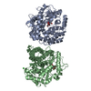
| |||||||||||||||||||||
|---|---|---|---|---|---|---|---|---|---|---|---|---|---|---|---|---|---|---|---|---|---|---|
| 1 | 
| |||||||||||||||||||||
| 2 | 
| |||||||||||||||||||||
| Unit cell |
| |||||||||||||||||||||
| Components on special symmetry positions |
|
-Components
| #1: Protein | Mass: 48839.117 Da / Num. of mol.: 2 Source method: isolated from a genetically manipulated source Source: (gene. exp.) Thermus thermophilus HB8 (bacteria) / Gene: TTHA0978 / Plasmid: pET-11a / Production host: #2: Chemical |   Mass: 35.453 Da / Num. of mol.: 2 / Source method: obtained synthetically / Formula: Cl Mass: 35.453 Da / Num. of mol.: 2 / Source method: obtained synthetically / Formula: Cl#3: Chemical |   Mass: 122.143 Da / Num. of mol.: 2 / Source method: obtained synthetically / Formula: C4H12NO3 / Comment: pH buffer*YM Mass: 122.143 Da / Num. of mol.: 2 / Source method: obtained synthetically / Formula: C4H12NO3 / Comment: pH buffer*YM#4: Chemical |   Mass: 106.077 Da / Num. of mol.: 2 / Source method: obtained synthetically / Formula: C3H6O4 Mass: 106.077 Da / Num. of mol.: 2 / Source method: obtained synthetically / Formula: C3H6O4#5: Water | ChemComp-HOH / |  Mass: 18.015 Da / Num. of mol.: 265 / Source method: isolated from a natural source / Formula: H2O Mass: 18.015 Da / Num. of mol.: 265 / Source method: isolated from a natural source / Formula: H2O |
|---|
-Experimental details
-Experiment
| Experiment | Method: X-RAY DIFFRACTION / Number of used crystals: 1 |
|---|
- Sample preparation
| Crystal | Density Matthews: 2.43 Å3/Da / Density % sol: 49.48 % |
|---|---|
| Crystal grow | Temperature: 293 K / Method: vapor diffusion, hanging drop / pH: 7.5 Details: 0.4M potassium sodium tartrate, 0.1M HEPES-NaOH (pH 7.5), 50mM calcium chloride, 10mM D-glycerate |
-Data collection
| Diffraction | Mean temperature: 100 K |
|---|---|
| Diffraction source | Source: SYNCHROTRON / Site: Photon Factory  / Beamline: AR-NE3A / Wavelength: 1 Å / Beamline: AR-NE3A / Wavelength: 1 Å |
| Detector | Type: ADSC QUANTUM 270 / Detector: CCD / Date: Jun 14, 2013 |
| Radiation | Protocol: SINGLE WAVELENGTH / Monochromatic (M) / Laue (L): M / Scattering type: x-ray |
| Radiation wavelength | Wavelength: 1 Å / Relative weight: 1 |
| Reflection | Resolution: 2.1→50 Å / Num. obs: 55067 / % possible obs: 97.6 % / Redundancy: 4.6 % / Rmerge(I) obs: 0.11 / Net I/σ(I): 9.3 |
| Reflection shell | Resolution: 2.1→2.21 Å / Redundancy: 4.2 % / Rmerge(I) obs: 0.19 / Mean I/σ(I) obs: 5.5 / % possible all: 93.6 |
- Processing
| Software |
| ||||||||||||||||||||||||||||||||||||||||||||||||||||||||||||||||||||||||||||||||||||||||||||||||||||||||||||||||||||||||||||||||||||||||||||||||||||||||||||||||||||||||||||||||||||||
|---|---|---|---|---|---|---|---|---|---|---|---|---|---|---|---|---|---|---|---|---|---|---|---|---|---|---|---|---|---|---|---|---|---|---|---|---|---|---|---|---|---|---|---|---|---|---|---|---|---|---|---|---|---|---|---|---|---|---|---|---|---|---|---|---|---|---|---|---|---|---|---|---|---|---|---|---|---|---|---|---|---|---|---|---|---|---|---|---|---|---|---|---|---|---|---|---|---|---|---|---|---|---|---|---|---|---|---|---|---|---|---|---|---|---|---|---|---|---|---|---|---|---|---|---|---|---|---|---|---|---|---|---|---|---|---|---|---|---|---|---|---|---|---|---|---|---|---|---|---|---|---|---|---|---|---|---|---|---|---|---|---|---|---|---|---|---|---|---|---|---|---|---|---|---|---|---|---|---|---|---|---|---|---|
| Refinement | Resolution: 2.1→45.145 Å / Cor.coef. Fo:Fc: 0.879 / Cor.coef. Fo:Fc free: 0.825 / Cross valid method: THROUGHOUT / ESU R: 0.287 / ESU R Free: 0.222 / Stereochemistry target values: MAXIMUM LIKELIHOOD / Details: HYDROGENS HAVE BEEN ADDED IN THE RIDING POSITIONS
| ||||||||||||||||||||||||||||||||||||||||||||||||||||||||||||||||||||||||||||||||||||||||||||||||||||||||||||||||||||||||||||||||||||||||||||||||||||||||||||||||||||||||||||||||||||||
| Solvent computation | Ion probe radii: 0.8 Å / Shrinkage radii: 0.8 Å / VDW probe radii: 1.2 Å / Solvent model: BABINET MODEL WITH MASK | ||||||||||||||||||||||||||||||||||||||||||||||||||||||||||||||||||||||||||||||||||||||||||||||||||||||||||||||||||||||||||||||||||||||||||||||||||||||||||||||||||||||||||||||||||||||
| Displacement parameters | Biso mean: 30.159 Å2
| ||||||||||||||||||||||||||||||||||||||||||||||||||||||||||||||||||||||||||||||||||||||||||||||||||||||||||||||||||||||||||||||||||||||||||||||||||||||||||||||||||||||||||||||||||||||
| Refinement step | Cycle: LAST / Resolution: 2.1→45.145 Å
| ||||||||||||||||||||||||||||||||||||||||||||||||||||||||||||||||||||||||||||||||||||||||||||||||||||||||||||||||||||||||||||||||||||||||||||||||||||||||||||||||||||||||||||||||||||||
| Refine LS restraints |
|