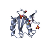
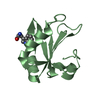
- PDB-6hip: Structure of SPF45 UHM bound to HIV-1 Rev ULM -
+
Open data
ID or keywords:
Loading...
-
Basic information
Entry
Database: PDB / ID: 6hip
Title
Structure of SPF45 UHM bound to HIV-1 Rev ULM
Components
HIV-1 Rev (41-49)
Splicing factor 45
Keywords
SPLICING / Splicing factor / viral protein
Function / homology
Function and homology information
host cell nucleolus / alternative mRNA splicing, via spliceosome / Rev-mediated nuclear export of HIV RNA / Nuclear import of Rev protein / mRNA cis splicing, via spliceosome / Integration of viral DNA into host genomic DNA / Autointegration results in viral DNA circles / Minus-strand DNA synthesis / Plus-strand DNA synthesis / Uncoating of the HIV Virion ...host cell nucleolus / alternative mRNA splicing, via spliceosome / Rev-mediated nuclear export of HIV RNA / Nuclear import of Rev protein / mRNA cis splicing, via spliceosome / Integration of viral DNA into host genomic DNA / Autointegration results in viral DNA circles / Minus-strand DNA synthesis / Plus-strand DNA synthesis / Uncoating of the HIV Virion / 2-LTR circle formation / Vpr-mediated nuclear import of PICs / Early Phase of HIV Life Cycle / Integration of provirus / APOBEC3G mediated resistance to HIV-1 infection / viral process / Binding and entry of HIV virion / mRNA transport / mRNA Splicing - Major Pathway / spliceosomal complex / Assembly Of The HIV Virion / Budding and maturation of HIV virion / host cell cytoplasm / DNA-binding transcription factor activity / RNA binding / nucleoplasm / identical protein binding Similarity search - Function
Resolution: 1.2→18.49 Å / Cor.coef. Fo:Fc: 0.959 / Cor.coef. Fo:Fc free: 0.947 / SU B: 1.15 / SU ML: 0.024 / Cross valid method: THROUGHOUT / ESU R: 0.042 / ESU R Free: 0.041 / Details: HYDROGENS HAVE BEEN ADDED IN THE RIDING POSITIONS
Rfactor
Num. reflection
% reflection
Selection details
Rfree
0.17502
3242
5.1 %
RANDOM
Rwork
0.14893
-
-
-
obs
0.15022
60832
97.41 %
-
Solvent computation
Ion probe radii: 0.8 Å / Shrinkage radii: 0.8 Å / VDW probe radii: 1.2 Å
 Movie
Movie Controller


 Open data
 Basic information
 Components
 Keywords
 Function and homology information
 Homo sapiens (human)
 Human immunodeficiency virus 1
 X-RAY DIFFRACTION /
  Authors
 Germany, 1items
  Citation
 Structure visualization
 Downloads & links
 Other downloads
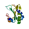


 PDBj
PDBj


 Assembly


 Type: L-peptide linking / Mass: 204.225 Da / Num. of mol.: 1 / Source method: obtained synthetically / Formula: C11H12N2O2
 Mass: 22.990 Da / Num. of mol.: 1 / Source method: obtained synthetically / Formula: Na
 Mass: 18.015 Da / Num. of mol.: 323 / Source method: isolated from a natural source / Formula: H2O
 Sample preparation
 / Beamline: X06SA / Wavelength: 0.99984 Å
 Processing