 ムービー
ムービー コントローラー


+ データを開く

- 基本情報
基本情報
| 登録情報 | データベース: PDB / ID: 1oi0 | ||||||
|---|---|---|---|---|---|---|---|
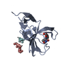
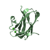
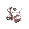
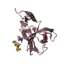
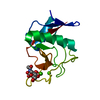
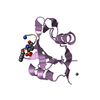
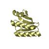
| タイトル | CRYSTAL STRUCTURE OF AF2198, A JAB1/MPN DOMAIN PROTEIN FROM ARCHAEOGLOBUS FULGIDUS | ||||||
 要素 要素 | HYPOTHETICAL PROTEIN AF2198 | ||||||
 キーワード キーワード | HYDROLASE / PROTEASOME / DEUBIQUITINATION / ARCHAEA | ||||||
| 機能・相同性 |  機能・相同性情報 機能・相同性情報 | ||||||
| 生物種 |   ARCHAEOGLOBUS FULGIDUS (古細菌) ARCHAEOGLOBUS FULGIDUS (古細菌) | ||||||
| 手法 |  X線回折 / シンクロトロン / 多重同系置換・異常分散 / 解像度: 1.5 Å X線回折 / シンクロトロン / 多重同系置換・異常分散 / 解像度: 1.5 Å | ||||||
 データ登録者 データ登録者 | Tran, H.J.T.T. / Allen, M.D. / Lowe, J. / Bycroft, M. | ||||||
 引用 引用 | ジャーナル: Biochemistry / 年: 2003 タイトル: The Structure of the Jab1/Mpn Domain and its Implications for Proteasome Function 著者: Tran, H.J.T.T. / Allen, M.D. / Lowe, J. / Bycroft, M. | ||||||
| 履歴 |
|
- 構造の表示
構造の表示
| 構造ビューア | 分子: MolmilJmol/JSmol |
|---|
- ダウンロードとリンク
-ダウンロード
| PDBx/mmCIF形式 | 1oi0.cif.gz | 102.3 KB | 表示 | PDBx/mmCIF形式 |
|---|---|---|---|---|
| PDB形式 | pdb1oi0.ent.gz | 79.1 KB | 表示 | PDB形式 |
| PDBx/mmJSON形式 | 1oi0.json.gz | ツリー表示 | PDBx/mmJSON形式 | |
| その他 |  その他のダウンロード その他のダウンロード |
-検証レポート
| アーカイブディレクトリ | https://data.pdbj.org/pub/pdb/validation_reports/oi/1oi0ftp://data.pdbj.org/pub/pdb/validation_reports/oi/1oi0 | HTTPS FTP |
|---|
-関連構造データ


-リンク
 PDBj
PDBj

- 集合体
集合体
| 登録構造単位 | 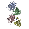
| ||||||||
|---|---|---|---|---|---|---|---|---|---|
| 1 | 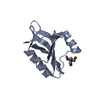
| ||||||||
| 2 | 
| ||||||||
| 3 | 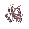
| ||||||||
| 4 | 
| ||||||||
| 単位格子 |
|
-要素
| #1: タンパク質 | 分子量: 13865.925 Da / 分子数: 4 / 由来タイプ: 組換発現 / 由来: (組換発現) ARCHAEOGLOBUS FULGIDUS (古細菌) / 発現宿主:  #2: 化合物 | ChemComp-ZN /   分子量: 65.409 Da / 分子数: 4 / 由来タイプ: 合成 / 式: Zn 分子量: 65.409 Da / 分子数: 4 / 由来タイプ: 合成 / 式: Zn#3: 化合物 |   分子量: 122.143 Da / 分子数: 2 / 由来タイプ: 合成 / 式: C4H12NO3 分子量: 122.143 Da / 分子数: 2 / 由来タイプ: 合成 / 式: C4H12NO3#4: 水 | ChemComp-HOH / |  分子量: 18.015 Da / 分子数: 278 / 由来タイプ: 天然 / 式: H2O 分子量: 18.015 Da / 分子数: 278 / 由来タイプ: 天然 / 式: H2OHas protein modification | Y | 配列の詳細 | ADDITIONAL | |
|---|
-実験情報
-実験
| 実験 | 手法: X線回折 / 使用した結晶の数: 1 |
|---|
- 試料調製
試料調製
| 結晶 | マシュー密度: 2.02 Å3/Da / 溶媒含有率: 39 % | |||||||||||||||||||||||||||||||||||||||||||||||||
|---|---|---|---|---|---|---|---|---|---|---|---|---|---|---|---|---|---|---|---|---|---|---|---|---|---|---|---|---|---|---|---|---|---|---|---|---|---|---|---|---|---|---|---|---|---|---|---|---|---|---|
| 結晶化 | pH: 7.8 詳細: 100MM MGCL2, 100MM TRIS (PH 7.8), 11% PEG 4000, 5 MM DTT | |||||||||||||||||||||||||||||||||||||||||||||||||
| 結晶化 | *PLUS 温度: 17 ℃ / pH: 7 / 手法: 蒸気拡散法, シッティングドロップ法 | |||||||||||||||||||||||||||||||||||||||||||||||||
| 溶液の組成 | *PLUS
|
-データ収集
| 回折 | 平均測定温度: 100 K |
|---|---|
| 放射光源 | 由来: シンクロトロン / サイト: ESRF  / ビームライン: ID14-1 / 波長: 0.979 / ビームライン: ID14-1 / 波長: 0.979 |
| 検出器 | タイプ: ADSC CCD / 検出器: CCD / 詳細: RIGAKU |
| 放射 | プロトコル: SINGLE WAVELENGTH / 単色(M)・ラウエ(L): M / 散乱光タイプ: x-ray |
| 放射波長 | 波長: 0.979 Å / 相対比: 1 |
| 反射 | 解像度: 1.5→25 Å / Num. obs: 69516 / % possible obs: 85.6 % / 冗長度: 3.5 % / Rmerge(I) obs: 0.067 / Net I/σ(I): 12.4 |
| 反射 シェル | 解像度: 1.5→1.58 Å / 冗長度: 3.6 % / Rmerge(I) obs: 0.338 / Mean I/σ(I) obs: 3.8 / % possible all: 78.6 |
| 反射 | *PLUS 最高解像度: 1.5 Å / 最低解像度: 25 Å / Rmerge(I) obs: 0.067 |
| 反射 シェル | *PLUS % possible obs: 78.6 % / Rmerge(I) obs: 0.338 |
- 解析
解析
| ソフトウェア |
| ||||||||||||||||||||||||||||||||||||||||||||||||||||||||||||
|---|---|---|---|---|---|---|---|---|---|---|---|---|---|---|---|---|---|---|---|---|---|---|---|---|---|---|---|---|---|---|---|---|---|---|---|---|---|---|---|---|---|---|---|---|---|---|---|---|---|---|---|---|---|---|---|---|---|---|---|---|---|
| 精密化 | 構造決定の手法: 多重同系置換・異常分散 / 解像度: 1.5→25 Å / 交差検証法: THROUGHOUT / σ(F): 0 / 詳細: DISORDERED REGIONS WERE NOT BUILT
| ||||||||||||||||||||||||||||||||||||||||||||||||||||||||||||
| 原子変位パラメータ | Biso mean: 24.98 Å2
| ||||||||||||||||||||||||||||||||||||||||||||||||||||||||||||
| 精密化ステップ | サイクル: LAST / 解像度: 1.5→25 Å
| ||||||||||||||||||||||||||||||||||||||||||||||||||||||||||||
| 拘束条件 |
| ||||||||||||||||||||||||||||||||||||||||||||||||||||||||||||
| LS精密化 シェル | 解像度: 1.5→1.51 Å / Num. reflection Rwork: 53 / Total num. of bins used: 50 | ||||||||||||||||||||||||||||||||||||||||||||||||||||||||||||
| 精密化 | *PLUS 最低解像度: 50 Å | ||||||||||||||||||||||||||||||||||||||||||||||||||||||||||||
| 溶媒の処理 | *PLUS | ||||||||||||||||||||||||||||||||||||||||||||||||||||||||||||
| 原子変位パラメータ | *PLUS |
