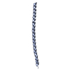
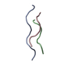
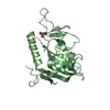
Entry Database : PDB / ID : 6hg7Title Crystal structure of a collagen II fragment containing the binding site of PEDF and COMP, (POG)4-LKG HRG FTG LQG-POG(4) Collagen alpha-1(II) chain Keywords / / / Function / homology Function Domain/homology Component
/ / / / / / / / / / / / / / / / / / / / / / / / / / / / / / / / / / / / / / / / / / / / / / / / / / / / / / / / / / / / / / / / / / / / / / / / / / / / / / / Biological species Homo sapiens (human)Method / / / Resolution : 1 Å Authors Gebauer, J.M. / Koehler, A. / Dietmar, H. / Gompert, M. / Neundorf, I. / Zaucke, F. / Koch, M. / Baumann, U. Funding support Organization Grant number Country German Research Foundation SFB829/B11
Journal : Sci Rep / Year : 2018Title : COMP and TSP-4 interact specifically with the novel GXKGHR motif only found in fibrillar collagens.Authors : Gebauer, J.M. / Kohler, A. / Dietmar, H. / Gompert, M. / Neundorf, I. / Zaucke, F. / Koch, M. / Baumann, U. History Deposition Aug 22, 2018 Deposition site / Processing site Revision 1.0 Dec 5, 2018 Provider / Type Revision 2.0 Jun 19, 2019 Group Atomic model / Data collection ... Atomic model / Data collection / Database references / Derived calculations / Non-polymer description / Polymer sequence / Source and taxonomy / Structure summary Category atom_site / atom_site_anisotrop ... atom_site / atom_site_anisotrop / chem_comp / entity / entity_name_com / entity_poly / entity_poly_seq / pdbx_entity_nonpoly / pdbx_entity_src_syn / pdbx_nonpoly_scheme / pdbx_poly_seq_scheme / pdbx_seq_map_depositor_info / pdbx_struct_assembly_gen / pdbx_struct_assembly_gen_depositor_info / pdbx_struct_mod_residue / struct_asym / struct_conn / struct_ref / struct_ref_seq / struct_ref_seq_dif / struct_site / struct_site_gen Item _atom_site.B_iso_or_equiv / _atom_site.Cartn_x ... _atom_site.B_iso_or_equiv / _atom_site.Cartn_x / _atom_site.Cartn_y / _atom_site.Cartn_z / _atom_site.auth_asym_id / _atom_site.auth_atom_id / _atom_site.auth_comp_id / _atom_site.auth_seq_id / _atom_site.group_PDB / _atom_site.label_alt_id / _atom_site.label_asym_id / _atom_site.label_atom_id / _atom_site.label_comp_id / _atom_site.label_entity_id / _atom_site.label_seq_id / _atom_site.occupancy / _atom_site.pdbx_auth_asym_id / _atom_site.pdbx_auth_atom_name / _atom_site.pdbx_auth_comp_id / _atom_site.pdbx_auth_seq_id / _atom_site.type_symbol / _atom_site_anisotrop.U[1][1] / _atom_site_anisotrop.U[1][2] / _atom_site_anisotrop.U[1][3] / _atom_site_anisotrop.U[2][2] / _atom_site_anisotrop.U[2][3] / _atom_site_anisotrop.U[3][3] / _atom_site_anisotrop.id / _atom_site_anisotrop.pdbx_PDB_atom_name / _atom_site_anisotrop.pdbx_PDB_residue_name / _atom_site_anisotrop.pdbx_PDB_residue_no / _atom_site_anisotrop.pdbx_PDB_strand_id / _atom_site_anisotrop.pdbx_auth_asym_id / _atom_site_anisotrop.pdbx_auth_atom_id / _atom_site_anisotrop.pdbx_auth_comp_id / _atom_site_anisotrop.pdbx_auth_seq_id / _atom_site_anisotrop.pdbx_label_alt_id / _atom_site_anisotrop.pdbx_label_asym_id / _atom_site_anisotrop.pdbx_label_atom_id / _atom_site_anisotrop.pdbx_label_comp_id / _atom_site_anisotrop.pdbx_label_seq_id / _atom_site_anisotrop.type_symbol / _chem_comp.formula / _chem_comp.formula_weight / _chem_comp.id / _chem_comp.mon_nstd_flag / _chem_comp.name / _chem_comp.pdbx_synonyms / _chem_comp.type / _entity_poly.pdbx_seq_one_letter_code / _entity_poly.pdbx_seq_one_letter_code_can / _pdbx_entity_src_syn.ncbi_taxonomy_id / _pdbx_entity_src_syn.organism_common_name / _pdbx_entity_src_syn.organism_scientific / _pdbx_entity_src_syn.pdbx_end_seq_num / _pdbx_seq_map_depositor_info.one_letter_code / _pdbx_seq_map_depositor_info.one_letter_code_mod / _pdbx_struct_assembly_gen.asym_id_list / _pdbx_struct_assembly_gen_depositor_info.asym_id_list / _struct_conn.ptnr2_auth_seq_id / _struct_conn.ptnr2_label_asym_id / _struct_conn.ptnr2_label_seq_id / _struct_ref.db_code / _struct_ref.db_name / _struct_ref.pdbx_align_begin / _struct_ref.pdbx_db_accession / _struct_ref.pdbx_seq_one_letter_code / _struct_ref_seq.db_align_beg / _struct_ref_seq.db_align_end / _struct_ref_seq.pdbx_auth_seq_align_end / _struct_ref_seq.pdbx_db_accession / _struct_ref_seq.seq_align_end Revision 2.1 Oct 16, 2019 Group / Category / Item Revision 2.2 Jan 17, 2024 Group / Database references / Refinement descriptionCategory chem_comp_atom / chem_comp_bond ... chem_comp_atom / chem_comp_bond / database_2 / pdbx_initial_refinement_model Item / _database_2.pdbx_database_accession
Show all Show less
 Movie
Movie Controller

 Yorodumi
 Open data
 Basic information
 Components
 Keywords
 Function and homology information
 Homo sapiens (human)
 X-RAY DIFFRACTION /
  Authors
 Germany, 1items
  Citation
 Structure visualization
 Downloads & links
 Other downloads
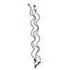


 PDBj
PDBj


 Assembly

 Mass: 96.063 Da / Num. of mol.: 1
 Mass: 18.015 Da / Num. of mol.: 164 / Source method: isolated from a natural source / Formula: H2O
 Sample preparation
 / Beamline: X06DA / Wavelength: 0.8 Å
 Processing