 Movie
Movie Controller

[English] 日本語
 Yorodumi
- PDB-6hg4: Crystal Structure of the human IL-17RC ECD in complex with human ... -
+ Open data

- Basic information
| Entry | Database: PDB / ID: 6hg4 | ||||||
|---|---|---|---|---|---|---|---|
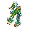
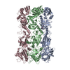
| Title | Crystal Structure of the human IL-17RC ECD in complex with human IL-17F | ||||||
 Components Components |
| ||||||
 Keywords Keywords | IMMUNE SYSTEM / Cystine-knot / FnIII domains / receptor-cytokine complex | ||||||
| Function / homology |  Function and homology information Function and homology informationregulation of granulocyte macrophage colony-stimulating factor production / regulation of interleukin-2 production / interleukin-17 receptor activity / positive regulation of lymphotoxin A production / granulocyte chemotaxis / regulation of interleukin-8 production / positive regulation of antimicrobial peptide production / Interleukin-17 signaling / interleukin-17A-mediated signaling pathway / regulation of transforming growth factor beta receptor signaling pathway ...regulation of granulocyte macrophage colony-stimulating factor production / regulation of interleukin-2 production / interleukin-17 receptor activity / positive regulation of lymphotoxin A production / granulocyte chemotaxis / regulation of interleukin-8 production / positive regulation of antimicrobial peptide production / Interleukin-17 signaling / interleukin-17A-mediated signaling pathway / regulation of transforming growth factor beta receptor signaling pathway / positive regulation of chemokine (C-X-C motif) ligand 1 production / interleukin-17-mediated signaling pathway / cytokine receptor binding / cartilage development / regulation of interleukin-6 production / positive regulation of cytokine production involved in inflammatory response / cytokine binding / defense response to fungus / coreceptor activity / negative regulation of angiogenesis / cytokine activity / positive regulation of cytokine production / positive regulation of interleukin-6 production / Interleukin-4 and Interleukin-13 signaling / defense response to Gram-negative bacterium / adaptive immune response / defense response to Gram-positive bacterium / inflammatory response / protein heterodimerization activity / signaling receptor binding / innate immune response / SARS-CoV-2 activates/modulates innate and adaptive immune responses / cell surface / protein homodimerization activity / positive regulation of transcription by RNA polymerase II / : / extracellular region / plasma membrane Similarity search - Function | ||||||
| Biological species |  Homo sapiens (human) Homo sapiens (human) | ||||||
| Method |  X-RAY DIFFRACTION / SYNCHROTRON / MOLECULAR REPLACEMENT / Resolution: 3.32 Å X-RAY DIFFRACTION / SYNCHROTRON / MOLECULAR REPLACEMENT / Resolution: 3.32 Å | ||||||
 Authors Authors | Rondeau, J.M. / Goepfert, A. | ||||||
 Citation Citation | Journal: Immunity / Year: 2020 Title: Structural Analysis Reveals that the Cytokine IL-17F Forms a Homodimeric Complex with Receptor IL-17RC to Drive IL-17RA-Independent Signaling. Authors: Goepfert, A. / Lehmann, S. / Blank, J. / Kolbinger, F. / Rondeau, J.M. | ||||||
| History |
|
- Structure visualization
| Structure viewer | Molecule: MolmilJmol/JSmol |
|---|
- Downloads & links
-Download
| PDBx/mmCIF format | 6hg4.cif.gz | 230.1 KB | Display | PDBx/mmCIF format |
|---|---|---|---|---|
| PDB format | pdb6hg4.ent.gz | 186.7 KB | Display | PDB format |
| PDBx/mmJSON format | 6hg4.json.gz | Tree view | PDBx/mmJSON format | |
| Others |  Other downloads Other downloads |
-Validation report
| Arichive directory | https://data.pdbj.org/pub/pdb/validation_reports/hg/6hg4ftp://data.pdbj.org/pub/pdb/validation_reports/hg/6hg4 | HTTPS FTP |
|---|
-Related structure data
| Related structure data |  6hg9C  6hgaC  6hgoC 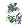 6hguC  1jpyS S: Starting model for refinement C: citing same article ( |
|---|---|
| Similar structure data |


-Links
 PDBj
PDBj

- Assembly
| Deposited unit | 
| ||||||||
|---|---|---|---|---|---|---|---|---|---|
| 1 | 
| ||||||||
| Unit cell |
|
-Components
| #1: Protein | Mass: 15695.895 Da / Num. of mol.: 1 / Fragment: IL-17F Source method: isolated from a genetically manipulated source Source: (gene. exp.) Homo sapiens (human) / Gene: IL17F / Plasmid: pCI-derived / Cell line (production host): HEK293S GnTi- / Production host: Homo sapiens (human) / References: UniProt: Q96PD4 |
|---|---|
| #2: Protein | Mass: 50454.312 Da / Num. of mol.: 1 / Mutation: N186Q, N226Q, N253Q, N263Q, N349Q, N372Q, N406Q Source method: isolated from a genetically manipulated source Details: Q307R variant with the following N-glycosylation site mutations: ,N186Q, N226Q, N253Q, N263Q, N349Q, N372Q, N406Q Source: (gene. exp.) Homo sapiens (human) / Gene: IL17RC, UNQ6118/PRO20040/PRO38901 / Plasmid: pCI-derived / Cell line (production host): HEK293S GnTi- / Production host: Homo sapiens (human) / References: UniProt: Q8NAC3 |
| Has protein modification | Y |
-Experimental details
-Experiment
| Experiment | Method: X-RAY DIFFRACTION / Number of used crystals: 1 |
|---|
- Sample preparation
| Crystal | Density Matthews: 4.09 Å3/Da / Density % sol: 69.96 % / Mosaicity: 0.21 ° |
|---|---|
| Crystal grow | Temperature: 293 K / Method: vapor diffusion, hanging drop / pH: 7 / Details: 200mM ammonium citrate tribasic, 20.0% PEG 3350 |
-Data collection
| Diffraction | Mean temperature: 100 K | ||||||||||||||||||||||||
|---|---|---|---|---|---|---|---|---|---|---|---|---|---|---|---|---|---|---|---|---|---|---|---|---|---|
| Diffraction source | Source: SYNCHROTRON / Site: SLS  / Beamline: X10SA / Wavelength: 0.99994 Å / Beamline: X10SA / Wavelength: 0.99994 Å | ||||||||||||||||||||||||
| Detector | Type: PSI PILATUS 6M / Detector: PIXEL / Date: Dec 12, 2016 / Details: mirrors | ||||||||||||||||||||||||
| Radiation | Protocol: SINGLE WAVELENGTH / Monochromatic (M) / Laue (L): M / Scattering type: x-ray | ||||||||||||||||||||||||
| Radiation wavelength | Wavelength: 0.99994 Å / Relative weight: 1 | ||||||||||||||||||||||||
| Reflection | Resolution: 3.32→133.66 Å / Num. obs: 16301 / % possible obs: 100 % / Observed criterion σ(I): -3 / Redundancy: 23.5 % / Biso Wilson estimate: 125.14 Å2 / CC1/2: 0.999 / Rmerge(I) obs: 0.181 / Rpim(I) all: 0.038 / Rrim(I) all: 0.185 / Net I/σ(I): 18 | ||||||||||||||||||||||||
| Reflection shell | Diffraction-ID: 1
|
- Processing
| Software |
| ||||||||||||||||||||||||||||||||||||||||||||||||||||||||||||||||||||||||||||||||||||||||||||||||||||||||||||||||||||||||||||||||||||||||||||||||||||||
|---|---|---|---|---|---|---|---|---|---|---|---|---|---|---|---|---|---|---|---|---|---|---|---|---|---|---|---|---|---|---|---|---|---|---|---|---|---|---|---|---|---|---|---|---|---|---|---|---|---|---|---|---|---|---|---|---|---|---|---|---|---|---|---|---|---|---|---|---|---|---|---|---|---|---|---|---|---|---|---|---|---|---|---|---|---|---|---|---|---|---|---|---|---|---|---|---|---|---|---|---|---|---|---|---|---|---|---|---|---|---|---|---|---|---|---|---|---|---|---|---|---|---|---|---|---|---|---|---|---|---|---|---|---|---|---|---|---|---|---|---|---|---|---|---|---|---|---|---|---|---|---|
| Refinement | Method to determine structure: MOLECULAR REPLACEMENT Starting model: 1jpy Resolution: 3.32→78.79 Å / Cor.coef. Fo:Fc: 0.921 / Cor.coef. Fo:Fc free: 0.853 / Cross valid method: THROUGHOUT / σ(F): 0 / SU Rfree Blow DPI: 0.391
| ||||||||||||||||||||||||||||||||||||||||||||||||||||||||||||||||||||||||||||||||||||||||||||||||||||||||||||||||||||||||||||||||||||||||||||||||||||||
| Displacement parameters | Biso max: 273.63 Å2 / Biso mean: 164.64 Å2 / Biso min: 78.74 Å2
| ||||||||||||||||||||||||||||||||||||||||||||||||||||||||||||||||||||||||||||||||||||||||||||||||||||||||||||||||||||||||||||||||||||||||||||||||||||||
| Refine analyze | Luzzati coordinate error obs: 0.44 Å | ||||||||||||||||||||||||||||||||||||||||||||||||||||||||||||||||||||||||||||||||||||||||||||||||||||||||||||||||||||||||||||||||||||||||||||||||||||||
| Refinement step | Cycle: final / Resolution: 3.32→78.79 Å
| ||||||||||||||||||||||||||||||||||||||||||||||||||||||||||||||||||||||||||||||||||||||||||||||||||||||||||||||||||||||||||||||||||||||||||||||||||||||
| Refine LS restraints |
| ||||||||||||||||||||||||||||||||||||||||||||||||||||||||||||||||||||||||||||||||||||||||||||||||||||||||||||||||||||||||||||||||||||||||||||||||||||||
| LS refinement shell | Resolution: 3.32→3.55 Å / Rfactor Rfree error: 0 / Total num. of bins used: 8
| ||||||||||||||||||||||||||||||||||||||||||||||||||||||||||||||||||||||||||||||||||||||||||||||||||||||||||||||||||||||||||||||||||||||||||||||||||||||
| Refinement TLS params. | Method: refined / Refine-ID: X-RAY DIFFRACTION
| ||||||||||||||||||||||||||||||||||||||||||||||||||||||||||||||||||||||||||||||||||||||||||||||||||||||||||||||||||||||||||||||||||||||||||||||||||||||
| Refinement TLS group |
|
