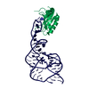
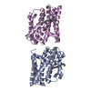
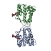
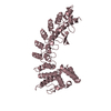
- PDB-6h56: Effector domain of Pseudomonas aeruginosa VgrG2b -
+
Open data
ID or keywords:
Loading...
-
Basic information
Entry
Database: PDB / ID: 6h56
Title
Effector domain of Pseudomonas aeruginosa VgrG2b
Components
Effector domain of Pseudomonas aeruginosa VgrG2b
Keywords
METAL BINDING PROTEIN / Zn binding / T6SS / effector
Function / homology
Function and homology information
protein secretion by the type VI secretion system / type VI protein secretion system complex / Hydrolases; Acting on peptide bonds (peptidases); Metalloendopeptidases / hydrolase activity / extracellular region / metal ion binding Similarity search - Function
Type VI secretion system spike protein VgrG2, C-terminal domain of unknown function DUF2345 / Putative type VI secretion system, Rhs element associated Vgr domain / Uncharacterized protein conserved in bacteria (DUF2345) / Putative type VI secretion system Rhs element Vgr / Type VI secretion system, RhsGE-associated Vgr family subset / Type VI secretion system, RhsGE-associated Vgr protein / Phage tail baseplate hub (GPD) / Gp5/Type VI secretion system Vgr protein, OB-fold domain / Type VI secretion system/phage-baseplate injector OB domain / Vgr protein, OB-fold domain superfamily ...Type VI secretion system spike protein VgrG2, C-terminal domain of unknown function DUF2345 / Putative type VI secretion system, Rhs element associated Vgr domain / Uncharacterized protein conserved in bacteria (DUF2345) / Putative type VI secretion system Rhs element Vgr / Type VI secretion system, RhsGE-associated Vgr family subset / Type VI secretion system, RhsGE-associated Vgr protein / Phage tail baseplate hub (GPD) / Gp5/Type VI secretion system Vgr protein, OB-fold domain / Type VI secretion system/phage-baseplate injector OB domain / Vgr protein, OB-fold domain superfamily / : / Neutral zinc metallopeptidases, zinc-binding region signature. Similarity search - Domain/homology
In the structure databanks used in Yorodumi, some data are registered as the other names, "COVID-19 virus" and "2019-nCoV". Here are the details of the virus and the list of structure data.
Jan 31, 2019. EMDB accession codes are about to change! (news from PDBe EMDB page)
EMDB accession codes are about to change! (news from PDBe EMDB page)
The allocation of 4 digits for EMDB accession codes will soon come to an end. Whilst these codes will remain in use, new EMDB accession codes will include an additional digit and will expand incrementally as the available range of codes is exhausted. The current 4-digit format prefixed with “EMD-” (i.e. EMD-XXXX) will advance to a 5-digit format (i.e. EMD-XXXXX), and so on. It is currently estimated that the 4-digit codes will be depleted around Spring 2019, at which point the 5-digit format will come into force.
The EM Navigator/Yorodumi systems omit the EMD- prefix.
Related info.:Q: What is EMD? / ID/Accession-code notation in Yorodumi/EM Navigator
Yorodumi is a browser for structure data from EMDB, PDB, SASBDB, etc.
This page is also the successor to EM Navigator detail page, and also detail information page/front-end page for Omokage search.
The word "yorodu" (or yorozu) is an old Japanese word meaning "ten thousand". "mi" (miru) is to see.
Related info.:EMDB / PDB / SASBDB / Comparison of 3 databanks / Yorodumi Search / Aug 31, 2016. New EM Navigator & Yorodumi / Yorodumi Papers / Jmol/JSmol / Function and homology information / Changes in new EM Navigator and Yorodumi
 Movie
Movie Controller


 Open data
 Basic information
 Components
 Keywords
 Function and homology information
 Pseudomonas aeruginosa PAO1 (bacteria)
 X-RAY DIFFRACTION /
  Authors
 United Kingdom, 1items
  Citation
 Structure visualization
 Downloads & links
 Other downloads
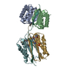


 PDBj
PDBj Assembly
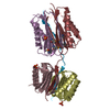

 Mass: 65.409 Da / Num. of mol.: 2 / Source method: obtained synthetically / Formula: Zn / Feature type: SUBJECT OF INVESTIGATION
 Sample preparation
 Processing