 Movie
Movie Controller


+ Open data

- Basic information
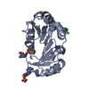
| Entry | Database: PDB / ID: 5o9j | ||||||||||||
|---|---|---|---|---|---|---|---|---|---|---|---|---|---|
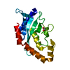
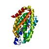
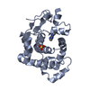
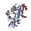
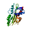
| Title | Crystal structure of transcription factor IIB Mja mini-intein | ||||||||||||
 Components Components | Transcription initiation factor IIB,Transcription initiation factor IIB | ||||||||||||
 Keywords Keywords | HYDROLASE / Transcription factor IIB / mini-intein / HINT fold | ||||||||||||
| Function / homology |  Function and homology information Function and homology informationtranscription preinitiation complex assembly / intron homing / intein-mediated protein splicing / transcription preinitiation complex / TBP-class protein binding / DNA-templated transcription initiation / endonuclease activity / Hydrolases; Acting on ester bonds / DNA-binding transcription factor activity / zinc ion binding Similarity search - Function | ||||||||||||
| Biological species |   Methanocaldococcus jannaschii (archaea) Methanocaldococcus jannaschii (archaea) | ||||||||||||
| Method |  X-RAY DIFFRACTION / SYNCHROTRON / MOLECULAR REPLACEMENT / Resolution: 2 Å X-RAY DIFFRACTION / SYNCHROTRON / MOLECULAR REPLACEMENT / Resolution: 2 Å | ||||||||||||
 Authors Authors | Mikula, K.M. / Iwai, H. / Zhou, D. / Wlodawer, A. | ||||||||||||
| Funding support |  Finland, Finland,  United States, 3items United States, 3items
| ||||||||||||
 Citation Citation | Journal: J. Mol. Biol. / Year: 2017 Title: Structural Basis for the Persistence of Homing Endonucleases in Transcription Factor IIB Inteins. Authors: Iwai, H. / Mikula, K.M. / Oeemig, J.S. / Zhou, D. / Li, M. / Wlodawer, A. | ||||||||||||
| History |
|
- Structure visualization
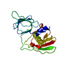
| Structure viewer | Molecule: MolmilJmol/JSmol |
|---|
- Downloads & links
-Download
| PDBx/mmCIF format | 5o9j.cif.gz | 95 KB | Display | PDBx/mmCIF format |
|---|---|---|---|---|
| PDB format | pdb5o9j.ent.gz | 71.1 KB | Display | PDB format |
| PDBx/mmJSON format | 5o9j.json.gz | Tree view | PDBx/mmJSON format | |
| Others |  Other downloads Other downloads |
-Validation report
| Arichive directory | https://data.pdbj.org/pub/pdb/validation_reports/o9/5o9jftp://data.pdbj.org/pub/pdb/validation_reports/o9/5o9j | HTTPS FTP |
|---|
-Related structure data
| Related structure data |  5o9iC  2cw8S S: Starting model for refinement C: citing same article ( |
|---|---|
| Similar structure data |

-Links
 PDBj
PDBj


- Assembly
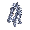
| Deposited unit | 
| ||||||||
|---|---|---|---|---|---|---|---|---|---|
| 1 | 
| ||||||||
| 2 | 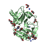
| ||||||||
| Unit cell |
|
-Components
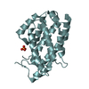
-Protein , 1 types, 2 molecules AB
| #1: Protein | Mass: 20872.121 Da / Num. of mol.: 2 Source method: isolated from a genetically manipulated source Source: (gene. exp.) Methanocaldococcus jannaschii (archaea)Strain: ATCC 43067 / DSM 2661 / JAL-1 / JCM 10045 / NBRC 100440 Gene: tfb, MJ0782 / Production host:  References: UniProt: Q58192, Hydrolases; Acting on ester bonds |
|---|
-Non-polymers , 5 types, 225 molecules 


| #2: Chemical | ChemComp-DIO /  Mass: 88.105 Da / Num. of mol.: 6 / Source method: obtained synthetically / Formula: C4H8O2 Mass: 88.105 Da / Num. of mol.: 6 / Source method: obtained synthetically / Formula: C4H8O2#3: Chemical | ChemComp-GOL /  Mass: 92.094 Da / Num. of mol.: 14 / Source method: obtained synthetically / Formula: C3H8O3 Mass: 92.094 Da / Num. of mol.: 14 / Source method: obtained synthetically / Formula: C3H8O3#4: Chemical | ChemComp-NH4 /  Mass: 18.038 Da / Num. of mol.: 10 / Source method: obtained synthetically / Formula: H4N Mass: 18.038 Da / Num. of mol.: 10 / Source method: obtained synthetically / Formula: H4N#5: Chemical | ChemComp-MES / |  Mass: 195.237 Da / Num. of mol.: 1 / Source method: obtained synthetically / Formula: C6H13NO4S / Comment: pH buffer*YM Mass: 195.237 Da / Num. of mol.: 1 / Source method: obtained synthetically / Formula: C6H13NO4S / Comment: pH buffer*YM#6: Water | ChemComp-HOH / | Mass: 18.015 Da / Num. of mol.: 194 / Source method: isolated from a natural source / Formula: H2O |
|---|
-Experimental details
-Experiment
| Experiment | Method: X-RAY DIFFRACTION / Number of used crystals: 1 |
|---|
- Sample preparation
| Crystal | Density Matthews: 2.88 Å3/Da / Density % sol: 57.34 % |
|---|---|
| Crystal grow | Temperature: 294 K / Method: vapor diffusion, sitting drop / pH: 6.5 / Details: MES, dioxane, ammonium sulfate |
-Data collection
| Diffraction | Mean temperature: 100 K |
|---|---|
| Diffraction source | Source: SYNCHROTRON / Site: Diamond  / Beamline: I04 / Wavelength: 0.9795 Å / Beamline: I04 / Wavelength: 0.9795 Å |
| Detector | Type: DECTRIS PILATUS 6M / Detector: PIXEL / Date: Jul 1, 2013 |
| Radiation | Protocol: SINGLE WAVELENGTH / Monochromatic (M) / Laue (L): M / Scattering type: x-ray |
| Radiation wavelength | Wavelength: 0.9795 Å / Relative weight: 1 |
| Reflection | Resolution: 2→28.7 Å / Num. obs: 30100 / % possible obs: 95.9 % / Redundancy: 2.4 % / Rmerge(I) obs: 0.085 / Net I/σ(I): 11.6 |
| Reflection shell | Highest resolution: 2.07 Å / Redundancy: 2.3 % / Rmerge(I) obs: 0.724 / Mean I/σ(I) obs: 1.6 / % possible all: 92.5 |
- Processing
| Software |
| ||||||||||||||||||||||||||||||||||||||||||||||||||||||||
|---|---|---|---|---|---|---|---|---|---|---|---|---|---|---|---|---|---|---|---|---|---|---|---|---|---|---|---|---|---|---|---|---|---|---|---|---|---|---|---|---|---|---|---|---|---|---|---|---|---|---|---|---|---|---|---|---|---|
| Refinement | Method to determine structure: MOLECULAR REPLACEMENT Starting model: 2CW8 Resolution: 2→28.7 Å / SU ML: 0.3 / Cross valid method: FREE R-VALUE / σ(F): 1.97 / Phase error: 33.92
| ||||||||||||||||||||||||||||||||||||||||||||||||||||||||
| Solvent computation | Shrinkage radii: 0.9 Å / VDW probe radii: 1.11 Å | ||||||||||||||||||||||||||||||||||||||||||||||||||||||||
| Refinement step | Cycle: LAST / Resolution: 2→28.7 Å
| ||||||||||||||||||||||||||||||||||||||||||||||||||||||||
| Refine LS restraints |
| ||||||||||||||||||||||||||||||||||||||||||||||||||||||||
| LS refinement shell |
|
