 Movie
Movie Controller


+ Open data

- Basic information
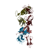
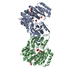
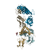
| Entry | Database: PDB / ID: 6gyz | ||||||
|---|---|---|---|---|---|---|---|
| Title | Crystal structure of GlmM from Staphylococcus aureus | ||||||
 Components Components | Phosphoglucosamine mutase | ||||||
 Keywords Keywords | ISOMERASE / cell wall / phosphoglucosamine mutase / peptidoglycan | ||||||
| Function / homology |  Function and homology information Function and homology informationphosphoglucosamine mutase / phosphoglucosamine mutase activity / carbohydrate metabolic process / magnesium ion binding Similarity search - Function | ||||||
| Biological species |   Staphylococcus aureus (bacteria) Staphylococcus aureus (bacteria) | ||||||
| Method |  X-RAY DIFFRACTION / SYNCHROTRON / MOLECULAR REPLACEMENT / Resolution: 3 Å X-RAY DIFFRACTION / SYNCHROTRON / MOLECULAR REPLACEMENT / Resolution: 3 Å | ||||||
 Authors Authors | Tosi, T. / Freemont, P.S. / Grundling, A. | ||||||
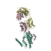
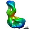
 Citation Citation | Journal: PLoS Pathog / Year: 2019 Title: Inhibition of the Staphylococcus aureus c-di-AMP cyclase DacA by direct interaction with the phosphoglucosamine mutase GlmM. Authors: Tommaso Tosi / Fumiya Hoshiga / Charlotte Millership / Rahul Singh / Charles Eldrid / Delphine Patin / Dominique Mengin-Lecreulx / Konstantinos Thalassinos / Paul Freemont / Angelika Gründling /   Abstract: c-di-AMP is an important second messenger molecule that plays a pivotal role in regulating fundamental cellular processes, including osmotic and cell wall homeostasis in many Gram-positive organisms. ...c-di-AMP is an important second messenger molecule that plays a pivotal role in regulating fundamental cellular processes, including osmotic and cell wall homeostasis in many Gram-positive organisms. In the opportunistic human pathogen Staphylococcus aureus, c-di-AMP is produced by the membrane-anchored DacA enzyme. Inactivation of this enzyme leads to a growth arrest under standard laboratory growth conditions and a re-sensitization of methicillin-resistant S. aureus (MRSA) strains to ß-lactam antibiotics. The gene coding for DacA is part of the conserved three-gene dacA/ybbR/glmM operon that also encodes the proposed DacA regulator YbbR and the essential phosphoglucosamine mutase GlmM, which is required for the production of glucosamine-1-phosphate, an early intermediate of peptidoglycan synthesis. These three proteins are thought to form a complex in vivo and, in this manner, help to fine-tune the cellular c-di-AMP levels. To further characterize this important regulatory complex, we conducted a comprehensive structural and functional analysis of the S. aureus DacA and GlmM enzymes by determining the structures of the S. aureus GlmM enzyme and the catalytic domain of DacA. Both proteins were found to be dimers in solution as well as in the crystal structures. Further site-directed mutagenesis, structural and enzymatic studies showed that multiple DacA dimers need to interact for enzymatic activity. We also show that DacA and GlmM form a stable complex in vitro and that S. aureus GlmM, but not Escherichia coli or Pseudomonas aeruginosa GlmM, acts as a strong inhibitor of DacA function without the requirement of any additional cellular factor. Based on Small Angle X-ray Scattering (SAXS) data, a model of the complex revealed that GlmM likely inhibits DacA by masking the active site of the cyclase and preventing higher oligomer formation. Together these results provide an important mechanistic insight into how c-di-AMP production can be regulated in the cell. | ||||||
| History |
|
- Structure visualization
| Structure viewer | Molecule: MolmilJmol/JSmol |
|---|
- Downloads & links
-Download
| PDBx/mmCIF format | 6gyz.cif.gz | 366.2 KB | Display | PDBx/mmCIF format |
|---|---|---|---|---|
| PDB format | pdb6gyz.ent.gz | 300.6 KB | Display | PDB format |
| PDBx/mmJSON format | 6gyz.json.gz | Tree view | PDBx/mmJSON format | |
| Others |  Other downloads Other downloads |
-Validation report
| Arichive directory | https://data.pdbj.org/pub/pdb/validation_reports/gy/6gyzftp://data.pdbj.org/pub/pdb/validation_reports/gy/6gyz | HTTPS FTP |
|---|
-Related structure data
| Related structure data | 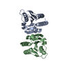 6gywC 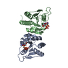 6gyxC 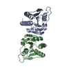 6gyyC  3pdkS S: Starting model for refinement C: citing same article ( |
|---|---|
| Similar structure data |


-Links
 PDBj
PDBj- Assembly
| Deposited unit | 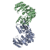
| ||||||||||||||||||||||||||||||||||||||||||||||||||||||||||||||||||||||||||||||||||
|---|---|---|---|---|---|---|---|---|---|---|---|---|---|---|---|---|---|---|---|---|---|---|---|---|---|---|---|---|---|---|---|---|---|---|---|---|---|---|---|---|---|---|---|---|---|---|---|---|---|---|---|---|---|---|---|---|---|---|---|---|---|---|---|---|---|---|---|---|---|---|---|---|---|---|---|---|---|---|---|---|---|---|---|
| 1 |
| ||||||||||||||||||||||||||||||||||||||||||||||||||||||||||||||||||||||||||||||||||
| Unit cell |
| ||||||||||||||||||||||||||||||||||||||||||||||||||||||||||||||||||||||||||||||||||
| Noncrystallographic symmetry (NCS) | NCS domain:
NCS domain segments: Ens-ID: 1
|