National Institutes of Health/National Institute of General Medical Sciences (NIH/NIGMS)
R35-GM128768
United States
Citation
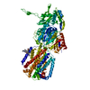
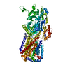
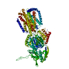
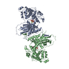
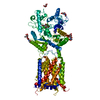
Journal: J Mol Biol / Year: 2021 Title: N-terminal Transmembrane-Helix Epitope Tag for X-ray Crystallography and Electron Microscopy of Small Membrane Proteins. Authors: Benjamin C McIlwain / Amanda L Erwin / Alexander R Davis / B Ben Koff / Louise Chang / Tatsiana Bylund / Gwo-Yu Chuang / Peter D Kwong / Melanie D Ohi / Yen-Ting Lai / Randy B Stockbridge / Abstract: Structural studies of membrane proteins, especially small membrane proteins, are associated with well-known experimental challenges. Complexation with monoclonal antibody fragments is a common ...Structural studies of membrane proteins, especially small membrane proteins, are associated with well-known experimental challenges. Complexation with monoclonal antibody fragments is a common strategy to augment such proteins; however, generating antibody fragments that specifically bind a target protein is not trivial. Here we identify a helical epitope, from the membrane-proximal external region (MPER) of the gp41-transmembrane subunit of the HIV envelope protein, that is recognized by several well-characterized antibodies and that can be fused as a contiguous extension of the N-terminal transmembrane helix of a broad range of membrane proteins. To analyze whether this MPER-epitope tag might aid structural studies of small membrane proteins, we determined an X-ray crystal structure of a membrane protein target that does not crystallize without the aid of crystallization chaperones, the Fluc fluoride channel, fused to the MPER epitope and in complex with antibody. We also demonstrate the utility of this approach for single particle electron microscopy with Fluc and two additional small membrane proteins that represent different membrane protein folds, AdiC and GlpF. These studies show that the MPER epitope provides a structurally defined, rigid docking site for antibody fragments that is transferable among diverse membrane proteins and can be engineered without prior structural information. Antibodies that bind to the MPER epitope serve as effective crystallization chaperones and electron microscopy fiducial markers, enabling structural studies of challenging small membrane proteins.
History
Deposition
Jan 6, 2021
-
Header (metadata) release
Mar 17, 2021
-
Map release
Mar 17, 2021
-
Update
Jul 28, 2021
-
Current status
Jul 28, 2021
Processing site: RCSB / Status: Released
-
Structure visualization
Movie
Surface view with section colored by density value
In the structure databanks used in Yorodumi, some data are registered as the other names, "COVID-19 virus" and "2019-nCoV". Here are the details of the virus and the list of structure data.
Jan 31, 2019. EMDB accession codes are about to change! (news from PDBe EMDB page)
EMDB accession codes are about to change! (news from PDBe EMDB page)
The allocation of 4 digits for EMDB accession codes will soon come to an end. Whilst these codes will remain in use, new EMDB accession codes will include an additional digit and will expand incrementally as the available range of codes is exhausted. The current 4-digit format prefixed with “EMD-” (i.e. EMD-XXXX) will advance to a 5-digit format (i.e. EMD-XXXXX), and so on. It is currently estimated that the 4-digit codes will be depleted around Spring 2019, at which point the 5-digit format will come into force.
The EM Navigator/Yorodumi systems omit the EMD- prefix.
Related info.:Q: What is EMD? / ID/Accession-code notation in Yorodumi/EM Navigator
Yorodumi is a browser for structure data from EMDB, PDB, SASBDB, etc.
This page is also the successor to EM Navigator detail page, and also detail information page/front-end page for Omokage search.
The word "yorodu" (or yorozu) is an old Japanese word meaning "ten thousand". "mi" (miru) is to see.
Related info.:EMDB / PDB / SASBDB / Comparison of 3 databanks / Yorodumi Search / Aug 31, 2016. New EM Navigator & Yorodumi / Yorodumi Papers / Jmol/JSmol / Function and homology information / Changes in new EM Navigator and Yorodumi
 Movie
Movie Controller


 Open data
 Basic information
 Map data
 Sample
 Bordetella pertussis (bacteria)
 Authors
 United States, 1 items
  Citation
 Structure visualization
 Movie viewer
 Downloads & links
 emd_23247.png
 http://ftp.pdbj.org/pub/emdb/structures/EMD-23247


 Z (Sec.)
 Y (Row.)
 X (Col.)


 Sample components
 Processing
 Electron microscopy
 FIELD EMISSION GUN

