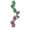
 Movie
Movie Controller


+ Open data

- Basic information
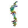
| Entry | Database: PDB / ID: 6x58 | ||||||
|---|---|---|---|---|---|---|---|
| Title | MPER-Fluc-Ec2 bound to 10E8v4 antibody | ||||||
 Components Components |
| ||||||
 Keywords Keywords | MEMBRANE PROTEIN / antibody / chaperone / MEMBRANE PROTEIN-IMMUNE SYSTEM complex | ||||||
| Function / homology |  Function and homology information Function and homology informationsymbiont-mediated suppression of host complement activation by recruitment of complement control protein / fluoride channel activity / cellular detoxification of fluoride / positive regulation of plasma membrane raft polarization / positive regulation of receptor clustering / host cell endosome membrane / clathrin-dependent endocytosis of virus by host cell / viral protein processing / fusion of virus membrane with host plasma membrane / fusion of virus membrane with host endosome membrane ...symbiont-mediated suppression of host complement activation by recruitment of complement control protein / fluoride channel activity / cellular detoxification of fluoride / positive regulation of plasma membrane raft polarization / positive regulation of receptor clustering / host cell endosome membrane / clathrin-dependent endocytosis of virus by host cell / viral protein processing / fusion of virus membrane with host plasma membrane / fusion of virus membrane with host endosome membrane / viral envelope / virion attachment to host cell / host cell plasma membrane / virion membrane / structural molecule activity / membrane / metal ion binding / plasma membrane Similarity search - Function | ||||||
| Biological species |  Homo sapiens (human) Homo sapiens (human)  Human immunodeficiency virus Human immunodeficiency virus | ||||||
| Method |  X-RAY DIFFRACTION / SYNCHROTRON / MOLECULAR REPLACEMENT / Resolution: 3.26 Å X-RAY DIFFRACTION / SYNCHROTRON / MOLECULAR REPLACEMENT / Resolution: 3.26 Å | ||||||
 Authors Authors | McIlwain, B.C. / Stockbridge, R.B. | ||||||
| Funding support |  United States, 1items United States, 1items
| ||||||
 Citation Citation | Journal: J Mol Biol / Year: 2021 Title: N-terminal Transmembrane-Helix Epitope Tag for X-ray Crystallography and Electron Microscopy of Small Membrane Proteins. Authors: Benjamin C McIlwain / Amanda L Erwin / Alexander R Davis / B Ben Koff / Louise Chang / Tatsiana Bylund / Gwo-Yu Chuang / Peter D Kwong / Melanie D Ohi / Yen-Ting Lai / Randy B Stockbridge / Abstract: Structural studies of membrane proteins, especially small membrane proteins, are associated with well-known experimental challenges. Complexation with monoclonal antibody fragments is a common ...Structural studies of membrane proteins, especially small membrane proteins, are associated with well-known experimental challenges. Complexation with monoclonal antibody fragments is a common strategy to augment such proteins; however, generating antibody fragments that specifically bind a target protein is not trivial. Here we identify a helical epitope, from the membrane-proximal external region (MPER) of the gp41-transmembrane subunit of the HIV envelope protein, that is recognized by several well-characterized antibodies and that can be fused as a contiguous extension of the N-terminal transmembrane helix of a broad range of membrane proteins. To analyze whether this MPER-epitope tag might aid structural studies of small membrane proteins, we determined an X-ray crystal structure of a membrane protein target that does not crystallize without the aid of crystallization chaperones, the Fluc fluoride channel, fused to the MPER epitope and in complex with antibody. We also demonstrate the utility of this approach for single particle electron microscopy with Fluc and two additional small membrane proteins that represent different membrane protein folds, AdiC and GlpF. These studies show that the MPER epitope provides a structurally defined, rigid docking site for antibody fragments that is transferable among diverse membrane proteins and can be engineered without prior structural information. Antibodies that bind to the MPER epitope serve as effective crystallization chaperones and electron microscopy fiducial markers, enabling structural studies of challenging small membrane proteins. | ||||||
| History |
|
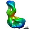
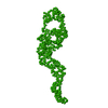
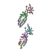
- Structure visualization
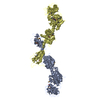
| Structure viewer | Molecule: MolmilJmol/JSmol |
|---|
- Downloads & links
-Download
| PDBx/mmCIF format | 6x58.cif.gz | 230.7 KB | Display | PDBx/mmCIF format |
|---|---|---|---|---|
| PDB format | pdb6x58.ent.gz | 185.1 KB | Display | PDB format |
| PDBx/mmJSON format | 6x58.json.gz | Tree view | PDBx/mmJSON format | |
| Others |  Other downloads Other downloads |
-Validation report
| Arichive directory | https://data.pdbj.org/pub/pdb/validation_reports/x5/6x58ftp://data.pdbj.org/pub/pdb/validation_reports/x5/6x58 | HTTPS FTP |
|---|
-Related structure data
| Related structure data | 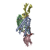 5a43S S: Starting model for refinement C: citing same article ( |
|---|---|
| Similar structure data |


-Links
 PDBj
PDBj


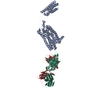
- Assembly
| Deposited unit | 
| ||||||||
|---|---|---|---|---|---|---|---|---|---|
| 1 |
| ||||||||
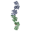
| Unit cell |
|
-Components
| #1: Antibody | Mass: 25138.002 Da / Num. of mol.: 2 Source method: isolated from a genetically manipulated source Source: (gene. exp.) Homo sapiens (human) / Cell line (production host): HEK / Production host: Homo sapiens (human) / Strain (production host): Expi293#2: Antibody | Mass: 22560.049 Da / Num. of mol.: 2 Source method: isolated from a genetically manipulated source Source: (gene. exp.) Homo sapiens (human) / Cell line (production host): HEK / Production host: Homo sapiens (human) / Strain (production host): Expi293#3: Protein | Mass: 15383.293 Da / Num. of mol.: 2 / Mutation: R25K,A51M in transporter Source method: isolated from a genetically manipulated source Source: (gene. exp.) Human immunodeficiency virus, (gene. exp.) Gene: env, crcB, crcB_2, flc_2, AC789_145pl00540, AKG29_01220, B6V57_25995, B7C53_24430, B9T59_30510, BANRA_05655, BANRA_05710, BEN53_26765, BIU72_24510, BK292_28205, BK334_22235, BK373_23795, BON66_ ...Gene: env, crcB, crcB_2, flc_2, AC789_145pl00540, AKG29_01220, B6V57_25995, B7C53_24430, B9T59_30510, BANRA_05655, BANRA_05710, BEN53_26765, BIU72_24510, BK292_28205, BK334_22235, BK373_23795, BON66_22385, BON69_17555, BON72_04410, BON76_23080, BON86_18360, BON94_16600, BVL39_28685, C2M16_27610, C4K41_18470, C4M78_26960, C5F73_28830, C5P01_26640, C5P44_26785, C6B13_25705, C7B08_22045, C9160_28010, C9201_28610, C9E25_24405, CDL37_21115, CSB64_25975, CWS33_26140, D1912_23150, D2184_26920, D2188_23810, D4U49_23430, D7W70_25855, D7Y10_22455, D9C99_23085, D9D33_24680, D9D94_24325, D9F87_24785, D9G11_25285, D9H70_24700, D9J44_26040, D9J61_22795, D9K02_24930, D9K48_12550, D9K54_12845, D9L99_22160, DAH18_26070, DB359_26980, DL545_01300, DLU67_24300, DLU82_24070, DN808_21420, DNI21_22025, DNQ45_14105, DNX19_24210, DP265_23725, DP277_24230, DQF72_24030, DS966_25260, DTL90_25845, DWB25_27055, E2112_25200, E2148_25435, EA239_25270, EA250_26195, EA429_25950, EA435_26495, ECONIH1_26550, ECS286_0026, EIA08_25210, EIZ93_19245, EJ366_00165, ELT22_23900, ELT58_23370, ELX56_24555, ELX56_26650, ELY05_20840, ELY05_25530, EPS76_20210, EVY14_24275, ExPECSC038_03841, EXX06_27990, EXX23_23730, EXX55_27910, EXX87_27755, EYX82_20275, F0L67_16895, F1E19_23340, F9050_24150, FORC82_p488, FQU83_00900, G3565_28150, GP698_23185, GQE58_24285, HVW93_24610, HXS78_24215, MJ49_27125, NCTC9001_00147, NCTC9077_06349, NCTC9434_05106, NCTC9969_05488, pCTXM15_EC8_00123, pO103_22, RCS79_P0115, SAMEA3472090_04449, SAMEA3752620_04806, SAMEA3753164_04868, SAMEA4370473_00090, TUM18780_48590, WP2S18E08_P10360, WP5S18E08_P10940 Production host: Has protein modification | Y | |
|---|
-Experimental details
-Experiment
| Experiment | Method: X-RAY DIFFRACTION / Number of used crystals: 1 |
|---|
- Sample preparation
| Crystal | Density Matthews: 3.8 Å3/Da / Density % sol: 67.66 % |
|---|---|
| Crystal grow | Temperature: 294 K / Method: vapor diffusion, sitting drop / Details: 34-39% PEG 300, 0.1 M NaCl, 0.1 M MES pH 6.2-7 / PH range: 6.2-7 |
-Data collection
| Diffraction | Mean temperature: 100 K / Serial crystal experiment: N | ||||||||||||||||||||||||||||||
|---|---|---|---|---|---|---|---|---|---|---|---|---|---|---|---|---|---|---|---|---|---|---|---|---|---|---|---|---|---|---|---|
| Diffraction source | Source: SYNCHROTRON / Site: APS / Beamline: 21-ID-D / Wavelength: 0.976 Å | ||||||||||||||||||||||||||||||
| Detector | Type: DECTRIS EIGER X 9M / Detector: PIXEL / Date: Jun 9, 2019 | ||||||||||||||||||||||||||||||
| Radiation | Protocol: SINGLE WAVELENGTH / Monochromatic (M) / Laue (L): M / Scattering type: x-ray | ||||||||||||||||||||||||||||||
| Radiation wavelength | Wavelength: 0.976 Å / Relative weight: 1 | ||||||||||||||||||||||||||||||
| Reflection | Resolution: 3.26→38.18 Å / Num. obs: 28368 / % possible obs: 98.6 % / Redundancy: 10.8 % / CC1/2: 0.999 / Rmerge(I) obs: 0.135 / Rpim(I) all: 0.043 / Rrim(I) all: 0.141 / Net I/σ(I): 10.3 / Num. measured all: 306181 / Scaling rejects: 3 | ||||||||||||||||||||||||||||||
| Reflection shell | Diffraction-ID: 1
|
- Processing
| Software |
| ||||||||||||||||||||
|---|---|---|---|---|---|---|---|---|---|---|---|---|---|---|---|---|---|---|---|---|---|
| Refinement | Method to determine structure: MOLECULAR REPLACEMENT Starting model: 5A43 Resolution: 3.26→34.02 Å / SU ML: 0.68 / Cross valid method: THROUGHOUT / σ(F): 1.96 / Phase error: 39.74 / Stereochemistry target values: ML
| ||||||||||||||||||||
| Solvent computation | Shrinkage radii: 0.9 Å / VDW probe radii: 1.11 Å / Solvent model: FLAT BULK SOLVENT MODEL | ||||||||||||||||||||
| Displacement parameters | Biso max: 322.54 Å2 / Biso mean: 159.7412 Å2 / Biso min: 30 Å2 | ||||||||||||||||||||
| Refinement step | Cycle: final / Resolution: 3.26→34.02 Å
|
