 Movie
Movie Controller


+ Open data

- Basic information
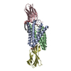
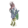
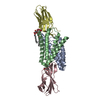
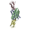
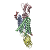
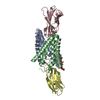
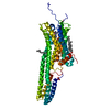
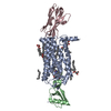
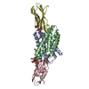
| Entry | Database: PDB / ID: 5a43 | ||||||
|---|---|---|---|---|---|---|---|
| Title | Crystal structure of a dual topology fluoride ion channel. | ||||||
 Components Components |
| ||||||
 Keywords Keywords | TRANSPORT PROTEIN / FLUORIDE ION CHANNEL / FLUC / EC2 | ||||||
| Function / homology |  Function and homology information Function and homology informationfluoride channel activity / cellular detoxification of fluoride / metal ion binding / plasma membrane Similarity search - Function | ||||||
| Biological species |   HOMO SAPIENS (human) HOMO SAPIENS (human) | ||||||
| Method |  X-RAY DIFFRACTION / SYNCHROTRON / SAD / Resolution: 2.58 Å X-RAY DIFFRACTION / SYNCHROTRON / SAD / Resolution: 2.58 Å | ||||||
 Authors Authors | Stockbridge, R.B. / Kolmakova-Partensky, L. / Shane, T. / Koide, A. / Koide, S. / Miller, C. / Newstead, S. | ||||||
 Citation Citation | Journal: Nature / Year: 2015 Title: Crystal Structures of a Double-Barrelled Fluoride Ion Channel. Authors: Stockbridge, R.B. / Kolmakova-Partensky, L. / Shane, T. / Koide, A. / Koide, S. / Miller, C. / Newstead, S. | ||||||
| History |
|
- Structure visualization
| Structure viewer | Molecule: MolmilJmol/JSmol |
|---|
- Downloads & links
-Download
| PDBx/mmCIF format | 5a43.cif.gz | 99.2 KB | Display | PDBx/mmCIF format |
|---|---|---|---|---|
| PDB format | pdb5a43.ent.gz | 76.9 KB | Display | PDB format |
| PDBx/mmJSON format | 5a43.json.gz | Tree view | PDBx/mmJSON format | |
| Others |  Other downloads Other downloads |
-Validation report
| Arichive directory | https://data.pdbj.org/pub/pdb/validation_reports/a4/5a43ftp://data.pdbj.org/pub/pdb/validation_reports/a4/5a43 | HTTPS FTP |
|---|
-Related structure data
| Related structure data | 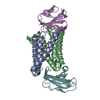 5a40C 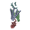 5nkqC  5a41 C: citing same article ( |
|---|---|
| Similar structure data |
-Links
 PDBj
PDBj- Assembly
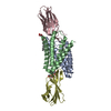
| Deposited unit | 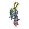
| ||||||||||||
|---|---|---|---|---|---|---|---|---|---|---|---|---|---|
| 1 |
| ||||||||||||
| 2 |
| ||||||||||||
| Unit cell |
| ||||||||||||
| Noncrystallographic symmetry (NCS) | NCS oper:
|
-Components
-Protein , 2 types, 4 molecules ABCD
| #1: Protein | Mass: 13863.006 Da / Num. of mol.: 2 Source method: isolated from a genetically manipulated source Source: (gene. exp.) #2: Protein | Mass: 10365.372 Da / Num. of mol.: 2 Source method: isolated from a genetically manipulated source Source: (gene. exp.) HOMO SAPIENS (human) / Plasmid: PHFT2 / Production host: |
|---|
-Sugars , 1 types, 3 molecules 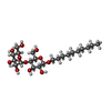
| #4: Sugar |  Type: D-saccharide / Mass: 482.562 Da / Num. of mol.: 3 Type: D-saccharide / Mass: 482.562 Da / Num. of mol.: 3Source method: isolated from a genetically manipulated source Formula: C22H42O11 / Comment: detergent*YM |
|---|
-Non-polymers , 3 types, 22 molecules 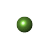


| #3: Chemical |  Mass: 18.998 Da / Num. of mol.: 2 / Source method: obtained synthetically / Formula: F Mass: 18.998 Da / Num. of mol.: 2 / Source method: obtained synthetically / Formula: F#5: Chemical | ChemComp-NA / |  Mass: 22.990 Da / Num. of mol.: 1 / Source method: obtained synthetically / Formula: Na Mass: 22.990 Da / Num. of mol.: 1 / Source method: obtained synthetically / Formula: Na#6: Water | ChemComp-HOH / | Mass: 18.015 Da / Num. of mol.: 19 / Source method: isolated from a natural source / Formula: H2O |
|---|
-Details
| Has protein modification | Y |
|---|
-Experimental details
-Experiment
| Experiment | Method: X-RAY DIFFRACTION / Number of used crystals: 1 |
|---|
- Sample preparation
| Crystal | Density Matthews: 6.03 Å3/Da / Density % sol: 79 % / Description: NONE |
|---|---|
| Crystal grow | pH: 6 / Details: 29% PEG550 MME, 50 MM LINO3, 100 MM ADA PH 6.0 |
-Data collection
| Diffraction | Mean temperature: 100 K |
|---|---|
| Diffraction source | Source: SYNCHROTRON / Site: ALS  / Beamline: 8.2.2 / Wavelength: 0.979 / Beamline: 8.2.2 / Wavelength: 0.979 |
| Detector | Type: ADSC QUANTUM 315 / Detector: CCD / Date: Nov 20, 2014 |
| Radiation | Protocol: SINGLE WAVELENGTH / Monochromatic (M) / Laue (L): M / Scattering type: x-ray |
| Radiation wavelength | Wavelength: 0.979 Å / Relative weight: 1 |
| Reflection | Resolution: 2.6→25 Å / Num. obs: 34593 / % possible obs: 99.9 % / Observed criterion σ(I): 2 / Redundancy: 14.3 % / Biso Wilson estimate: 62.65 Å2 / Rmerge(I) obs: 0.17 / Net I/σ(I): 9 |
| Reflection shell | Resolution: 2.6→2.7 Å / Redundancy: 14.3 % / Rmerge(I) obs: 0.99 / Mean I/σ(I) obs: 1.9 / % possible all: 100 |
- Processing
| Software |
| ||||||||||||||||||||||||||||||||||||||||||||||||||||||||||||||||||||||||||||||||||||||||||||||||||
|---|---|---|---|---|---|---|---|---|---|---|---|---|---|---|---|---|---|---|---|---|---|---|---|---|---|---|---|---|---|---|---|---|---|---|---|---|---|---|---|---|---|---|---|---|---|---|---|---|---|---|---|---|---|---|---|---|---|---|---|---|---|---|---|---|---|---|---|---|---|---|---|---|---|---|---|---|---|---|---|---|---|---|---|---|---|---|---|---|---|---|---|---|---|---|---|---|---|---|---|
| Refinement | Method to determine structure: SAD Starting model: NONE Resolution: 2.58→24.246 Å / SU ML: 0.45 / σ(F): 1.36 / Phase error: 32.67 / Stereochemistry target values: ML
| ||||||||||||||||||||||||||||||||||||||||||||||||||||||||||||||||||||||||||||||||||||||||||||||||||
| Solvent computation | Shrinkage radii: 0.9 Å / VDW probe radii: 1.11 Å / Solvent model: FLAT BULK SOLVENT MODEL | ||||||||||||||||||||||||||||||||||||||||||||||||||||||||||||||||||||||||||||||||||||||||||||||||||
| Displacement parameters | Biso mean: 62.65 Å2 | ||||||||||||||||||||||||||||||||||||||||||||||||||||||||||||||||||||||||||||||||||||||||||||||||||
| Refinement step | Cycle: LAST / Resolution: 2.58→24.246 Å
| ||||||||||||||||||||||||||||||||||||||||||||||||||||||||||||||||||||||||||||||||||||||||||||||||||
| Refine LS restraints |
| ||||||||||||||||||||||||||||||||||||||||||||||||||||||||||||||||||||||||||||||||||||||||||||||||||
| LS refinement shell |
|
