 Movie
Movie Controller

[English] 日本語
 Yorodumi
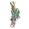
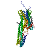
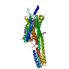
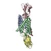
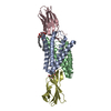
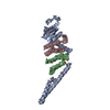
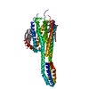
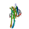
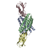
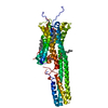
- PDB-4xt1: Structure of a nanobody-bound viral GPCR bound to human chemokine... -
+ Open data

- Basic information
| Entry | Database: PDB / ID: 4xt1 | |||||||||
|---|---|---|---|---|---|---|---|---|---|---|
| Title | Structure of a nanobody-bound viral GPCR bound to human chemokine CX3CL1 | |||||||||
 Components Components |
| |||||||||
 Keywords Keywords | VIRAL PROTEIN/SIGNALLING PROTEIN / GPCR / chemokine / membrane protein / complex / VIRAL PROTEIN-SIGNALLING PROTEIN complex | |||||||||
| Function / homology |  Function and homology information Function and homology informationCXCR1 chemokine receptor binding / positive regulation of calcium-independent cell-cell adhesion / negative regulation of interleukin-1 alpha production / leukocyte adhesive activation / CX3C chemokine receptor binding / negative regulation of glutamate receptor signaling pathway / lymphocyte chemotaxis / autocrine signaling / synapse pruning / positive regulation of microglial cell migration ...CXCR1 chemokine receptor binding / positive regulation of calcium-independent cell-cell adhesion / negative regulation of interleukin-1 alpha production / leukocyte adhesive activation / CX3C chemokine receptor binding / negative regulation of glutamate receptor signaling pathway / lymphocyte chemotaxis / autocrine signaling / synapse pruning / positive regulation of microglial cell migration / regulation of lipopolysaccharide-mediated signaling pathway / negative regulation of microglial cell activation / negative regulation of hippocampal neuron apoptotic process / negative regulation of neuron migration / positive regulation of transforming growth factor beta1 production / CCR chemokine receptor binding / microglial cell proliferation / leukocyte migration involved in inflammatory response / positive regulation of actin filament bundle assembly / integrin activation / C-C chemokine binding / angiogenesis involved in wound healing / eosinophil chemotaxis / C-C chemokine receptor activity / positive regulation of neuroblast proliferation / chemokine activity / Chemokine receptors bind chemokines / leukocyte chemotaxis / negative regulation of interleukin-1 beta production / positive regulation of cell-matrix adhesion / symbiont-mediated transformation of host cell / positive chemotaxis / neuron remodeling / chemoattractant activity / negative regulation of interleukin-6 production / macrophage chemotaxis / negative regulation of cell-substrate adhesion / negative regulation of apoptotic signaling pathway / negative regulation of tumor necrosis factor production / regulation of neurogenesis / negative regulation of extrinsic apoptotic signaling pathway in absence of ligand / extrinsic apoptotic signaling pathway in absence of ligand / cell projection / neutrophil chemotaxis / positive regulation of smooth muscle cell proliferation / response to ischemia / negative regulation of cell migration / positive regulation of release of sequestered calcium ion into cytosol / chemokine-mediated signaling pathway / calcium-mediated signaling / cell chemotaxis / neuron cellular homeostasis / defense response / positive regulation of neuron projection development / cell-cell adhesion / microglial cell activation / regulation of synaptic plasticity / integrin binding / chemotaxis / cytokine-mediated signaling pathway / positive regulation of angiogenesis / positive regulation of inflammatory response / cell-cell signaling / antimicrobial humoral immune response mediated by antimicrobial peptide / positive regulation of cytosolic calcium ion concentration / G alpha (i) signalling events / positive regulation of MAPK cascade / positive regulation of ERK1 and ERK2 cascade / positive regulation of canonical NF-kappaB signal transduction / positive regulation of phosphatidylinositol 3-kinase/protein kinase B signal transduction / cell adhesion / neuron projection / immune response / positive regulation of cell migration / inflammatory response / G protein-coupled receptor signaling pathway / signaling receptor binding / neuronal cell body / positive regulation of cell population proliferation / negative regulation of apoptotic process / host cell plasma membrane / perinuclear region of cytoplasm / cell surface / positive regulation of transcription by RNA polymerase II / : / extracellular region / membrane / plasma membrane Similarity search - Function | |||||||||
| Biological species |  Cytomegalovirus Cytomegalovirus Homo sapiens (human) Homo sapiens (human) | |||||||||
| Method |  X-RAY DIFFRACTION / SYNCHROTRON / MOLECULAR REPLACEMENT / Resolution: 2.886 Å X-RAY DIFFRACTION / SYNCHROTRON / MOLECULAR REPLACEMENT / Resolution: 2.886 Å | |||||||||
 Authors Authors | Burg, J.S. / Jude, K.M. / Waghray, D. / Garcia, K.C. | |||||||||
 Citation Citation | Journal: Science / Year: 2015 Title: Structural biology. Structural basis for chemokine recognition and activation of a viral G protein-coupled receptor. Authors: Burg, J.S. / Ingram, J.R. / Venkatakrishnan, A.J. / Jude, K.M. / Dukkipati, A. / Feinberg, E.N. / Angelini, A. / Waghray, D. / Dror, R.O. / Ploegh, H.L. / Garcia, K.C. | |||||||||
| History |
|
- Structure visualization
| Structure viewer | Molecule: MolmilJmol/JSmol |
|---|
- Downloads & links
-Download
| PDBx/mmCIF format | 4xt1.cif.gz | 217 KB | Display | PDBx/mmCIF format |
|---|---|---|---|---|
| PDB format | pdb4xt1.ent.gz | 171 KB | Display | PDB format |
| PDBx/mmJSON format | 4xt1.json.gz | Tree view | PDBx/mmJSON format | |
| Others |  Other downloads Other downloads |
-Validation report
| Arichive directory | https://data.pdbj.org/pub/pdb/validation_reports/xt/4xt1ftp://data.pdbj.org/pub/pdb/validation_reports/xt/4xt1 | HTTPS FTP |
|---|
-Related structure data
| Related structure data |  4xt3C  1f2lS  3onaS  4b41S 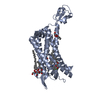 4mbsS C: citing same article ( S: Starting model for refinement |
|---|---|
| Similar structure data |
-Links
 PDBj
PDBj


- Assembly
| Deposited unit | 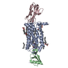
| ||||||||
|---|---|---|---|---|---|---|---|---|---|
| 1 |
| ||||||||
| Unit cell |
|
-Components
-Protein , 2 types, 2 molecules AB
| #1: Protein | Mass: 42041.098 Da / Num. of mol.: 1 Source method: isolated from a genetically manipulated source Source: (gene. exp.) Cytomegalovirus / Cell line (production host): HEK293s GnTI- / Production host: Homo sapiens (human) / References: UniProt: P69332 |
|---|---|
| #2: Protein | Mass: 9970.462 Da / Num. of mol.: 1 / Fragment: UNP residues 25-101 / Mutation: N9A Source method: isolated from a genetically manipulated source Source: (gene. exp.) Homo sapiens (human) / Gene: CX3CL1, FKN, NTT, SCYD1, A-152E5.2 / Cell line (production host): HEK293s GnTI- / Production host: Homo sapiens (human) / References: UniProt: P78423 |
-Antibody , 1 types, 1 molecules C
| #3: Antibody | Mass: 14624.158 Da / Num. of mol.: 1 Source method: isolated from a genetically manipulated source Source: (gene. exp.)  |
|---|
-Non-polymers , 5 types, 39 molecules 


| #4: Chemical |  Mass: 386.654 Da / Num. of mol.: 2 / Source method: obtained synthetically / Formula: C27H46O Mass: 386.654 Da / Num. of mol.: 2 / Source method: obtained synthetically / Formula: C27H46O#5: Chemical | ChemComp-OLC / (  Mass: 356.540 Da / Num. of mol.: 6 / Source method: obtained synthetically / Formula: C21H40O4 Mass: 356.540 Da / Num. of mol.: 6 / Source method: obtained synthetically / Formula: C21H40O4#6: Chemical | ChemComp-UNL / Num. of mol.: 7 / Source method: obtained synthetically #7: Chemical | ChemComp-SIN / |  Mass: 118.088 Da / Num. of mol.: 1 / Source method: obtained synthetically / Formula: C4H6O4 Mass: 118.088 Da / Num. of mol.: 1 / Source method: obtained synthetically / Formula: C4H6O4#8: Water | ChemComp-HOH / | Mass: 18.015 Da / Num. of mol.: 23 / Source method: isolated from a natural source / Formula: H2O |
|---|
-Details
| Has protein modification | Y |
|---|
-Experimental details
-Experiment
| Experiment | Method: X-RAY DIFFRACTION / Number of used crystals: 2 |
|---|
- Sample preparation
| Crystal | Density Matthews: 2.85 Å3/Da / Density % sol: 56.8 % |
|---|---|
| Crystal grow | Temperature: 293 K / Method: lipidic cubic phase / pH: 6.3 Details: PEG 300, MES, sodium succinate, polypropylene glycol 400 |
-Data collection
| Diffraction | Mean temperature: 100 K | |||||||||||||||||||||||||||||||||||||||||||||||||||||||||||||||||||||||||||||||||||||||||||||||||||||||||||||||||||||||||||||||||||||||||||||||||||||||||||||||||||||||||||||||||||||||||||||
|---|---|---|---|---|---|---|---|---|---|---|---|---|---|---|---|---|---|---|---|---|---|---|---|---|---|---|---|---|---|---|---|---|---|---|---|---|---|---|---|---|---|---|---|---|---|---|---|---|---|---|---|---|---|---|---|---|---|---|---|---|---|---|---|---|---|---|---|---|---|---|---|---|---|---|---|---|---|---|---|---|---|---|---|---|---|---|---|---|---|---|---|---|---|---|---|---|---|---|---|---|---|---|---|---|---|---|---|---|---|---|---|---|---|---|---|---|---|---|---|---|---|---|---|---|---|---|---|---|---|---|---|---|---|---|---|---|---|---|---|---|---|---|---|---|---|---|---|---|---|---|---|---|---|---|---|---|---|---|---|---|---|---|---|---|---|---|---|---|---|---|---|---|---|---|---|---|---|---|---|---|---|---|---|---|---|---|---|---|---|---|
| Diffraction source | Source: SYNCHROTRON / Site: APS  / Beamline: 23-ID-B / Wavelength: 1.033 Å / Beamline: 23-ID-B / Wavelength: 1.033 Å | |||||||||||||||||||||||||||||||||||||||||||||||||||||||||||||||||||||||||||||||||||||||||||||||||||||||||||||||||||||||||||||||||||||||||||||||||||||||||||||||||||||||||||||||||||||||||||||
| Detector | Type: MARMOSAIC 300 mm CCD / Detector: CCD / Date: Oct 13, 2013 | |||||||||||||||||||||||||||||||||||||||||||||||||||||||||||||||||||||||||||||||||||||||||||||||||||||||||||||||||||||||||||||||||||||||||||||||||||||||||||||||||||||||||||||||||||||||||||||
| Radiation | Protocol: SINGLE WAVELENGTH / Monochromatic (M) / Laue (L): M / Scattering type: x-ray | |||||||||||||||||||||||||||||||||||||||||||||||||||||||||||||||||||||||||||||||||||||||||||||||||||||||||||||||||||||||||||||||||||||||||||||||||||||||||||||||||||||||||||||||||||||||||||||
| Radiation wavelength | Wavelength: 1.033 Å / Relative weight: 1 | |||||||||||||||||||||||||||||||||||||||||||||||||||||||||||||||||||||||||||||||||||||||||||||||||||||||||||||||||||||||||||||||||||||||||||||||||||||||||||||||||||||||||||||||||||||||||||||
| Reflection | Resolution: 2.88→50 Å / Num. obs: 16648 / % possible obs: 98.7 % / Redundancy: 8.5 % / Biso Wilson estimate: 58.12 Å2 / Rmerge(I) obs: 0.2 / Rpim(I) all: 0.084 / Rrim(I) all: 0.214 / Χ2: 1.137 / Net I/av σ(I): 10.745 / Net I/σ(I): 5.2 / Num. measured all: 141947 | |||||||||||||||||||||||||||||||||||||||||||||||||||||||||||||||||||||||||||||||||||||||||||||||||||||||||||||||||||||||||||||||||||||||||||||||||||||||||||||||||||||||||||||||||||||||||||||
| Reflection shell | Diffraction-ID: 1 / Rejects: _
|
- Processing
| Software |
| |||||||||||||||||||||||||||||||||||||||||||||||||||||||||||||||||||||||||||||||||||||||||||||||||||||||||||||||||||||||||||||||||||||||||||||||||||||||||||||||||||||||||||||||||||||||||||||||||||||||||||||||||||||||||||||||||||||||||||||||||||||||||||||||||||||||||||||||||||||||||||||||||||||||||||||||||||||||||||||||||||||||||||||||||||||||||||||||||||||||||||||||||||||||
|---|---|---|---|---|---|---|---|---|---|---|---|---|---|---|---|---|---|---|---|---|---|---|---|---|---|---|---|---|---|---|---|---|---|---|---|---|---|---|---|---|---|---|---|---|---|---|---|---|---|---|---|---|---|---|---|---|---|---|---|---|---|---|---|---|---|---|---|---|---|---|---|---|---|---|---|---|---|---|---|---|---|---|---|---|---|---|---|---|---|---|---|---|---|---|---|---|---|---|---|---|---|---|---|---|---|---|---|---|---|---|---|---|---|---|---|---|---|---|---|---|---|---|---|---|---|---|---|---|---|---|---|---|---|---|---|---|---|---|---|---|---|---|---|---|---|---|---|---|---|---|---|---|---|---|---|---|---|---|---|---|---|---|---|---|---|---|---|---|---|---|---|---|---|---|---|---|---|---|---|---|---|---|---|---|---|---|---|---|---|---|---|---|---|---|---|---|---|---|---|---|---|---|---|---|---|---|---|---|---|---|---|---|---|---|---|---|---|---|---|---|---|---|---|---|---|---|---|---|---|---|---|---|---|---|---|---|---|---|---|---|---|---|---|---|---|---|---|---|---|---|---|---|---|---|---|---|---|---|---|---|---|---|---|---|---|---|---|---|---|---|---|---|---|---|---|---|---|---|---|---|---|---|---|---|---|---|---|---|---|---|---|---|---|---|---|---|---|---|---|---|---|---|---|---|---|---|---|---|---|---|---|---|---|---|---|---|---|---|---|---|---|---|---|---|---|---|---|---|---|---|---|---|---|---|---|---|---|---|---|---|---|---|---|---|---|---|---|---|---|---|---|---|---|---|---|---|---|---|---|---|---|---|---|---|---|---|---|---|---|---|---|---|---|---|---|---|
| Refinement | Method to determine structure: MOLECULAR REPLACEMENT Starting model: 4MBS, 1F2L, 3ONA, 4B41 Resolution: 2.886→38.234 Å / SU ML: 0.43 / Cross valid method: FREE R-VALUE / σ(F): 1.35 / Phase error: 27.33 / Stereochemistry target values: ML
| |||||||||||||||||||||||||||||||||||||||||||||||||||||||||||||||||||||||||||||||||||||||||||||||||||||||||||||||||||||||||||||||||||||||||||||||||||||||||||||||||||||||||||||||||||||||||||||||||||||||||||||||||||||||||||||||||||||||||||||||||||||||||||||||||||||||||||||||||||||||||||||||||||||||||||||||||||||||||||||||||||||||||||||||||||||||||||||||||||||||||||||||||||||||
| Solvent computation | Shrinkage radii: 0.9 Å / VDW probe radii: 1.11 Å / Solvent model: FLAT BULK SOLVENT MODEL | |||||||||||||||||||||||||||||||||||||||||||||||||||||||||||||||||||||||||||||||||||||||||||||||||||||||||||||||||||||||||||||||||||||||||||||||||||||||||||||||||||||||||||||||||||||||||||||||||||||||||||||||||||||||||||||||||||||||||||||||||||||||||||||||||||||||||||||||||||||||||||||||||||||||||||||||||||||||||||||||||||||||||||||||||||||||||||||||||||||||||||||||||||||||
| Displacement parameters | Biso max: 206.21 Å2 / Biso mean: 66.9984 Å2 / Biso min: 21.81 Å2 | |||||||||||||||||||||||||||||||||||||||||||||||||||||||||||||||||||||||||||||||||||||||||||||||||||||||||||||||||||||||||||||||||||||||||||||||||||||||||||||||||||||||||||||||||||||||||||||||||||||||||||||||||||||||||||||||||||||||||||||||||||||||||||||||||||||||||||||||||||||||||||||||||||||||||||||||||||||||||||||||||||||||||||||||||||||||||||||||||||||||||||||||||||||||
| Refinement step | Cycle: final / Resolution: 2.886→38.234 Å
| |||||||||||||||||||||||||||||||||||||||||||||||||||||||||||||||||||||||||||||||||||||||||||||||||||||||||||||||||||||||||||||||||||||||||||||||||||||||||||||||||||||||||||||||||||||||||||||||||||||||||||||||||||||||||||||||||||||||||||||||||||||||||||||||||||||||||||||||||||||||||||||||||||||||||||||||||||||||||||||||||||||||||||||||||||||||||||||||||||||||||||||||||||||||
| Refine LS restraints |
| |||||||||||||||||||||||||||||||||||||||||||||||||||||||||||||||||||||||||||||||||||||||||||||||||||||||||||||||||||||||||||||||||||||||||||||||||||||||||||||||||||||||||||||||||||||||||||||||||||||||||||||||||||||||||||||||||||||||||||||||||||||||||||||||||||||||||||||||||||||||||||||||||||||||||||||||||||||||||||||||||||||||||||||||||||||||||||||||||||||||||||||||||||||||
| LS refinement shell | Refine-ID: X-RAY DIFFRACTION / Total num. of bins used: 12
| |||||||||||||||||||||||||||||||||||||||||||||||||||||||||||||||||||||||||||||||||||||||||||||||||||||||||||||||||||||||||||||||||||||||||||||||||||||||||||||||||||||||||||||||||||||||||||||||||||||||||||||||||||||||||||||||||||||||||||||||||||||||||||||||||||||||||||||||||||||||||||||||||||||||||||||||||||||||||||||||||||||||||||||||||||||||||||||||||||||||||||||||||||||||
| Refinement TLS params. | Method: refined / Refine-ID: X-RAY DIFFRACTION
| |||||||||||||||||||||||||||||||||||||||||||||||||||||||||||||||||||||||||||||||||||||||||||||||||||||||||||||||||||||||||||||||||||||||||||||||||||||||||||||||||||||||||||||||||||||||||||||||||||||||||||||||||||||||||||||||||||||||||||||||||||||||||||||||||||||||||||||||||||||||||||||||||||||||||||||||||||||||||||||||||||||||||||||||||||||||||||||||||||||||||||||||||||||||
| Refinement TLS group |
|