 Movie
Movie Controller

[English] 日本語
 Yorodumi
- PDB-4b41: Crystal structure of an amyloid-beta binding single chain antibody G7 -
+ Open data

- Basic information
| Entry | Database: PDB / ID: 4b41 | ||||||
|---|---|---|---|---|---|---|---|
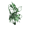
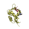
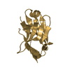
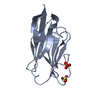
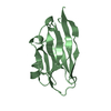
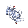
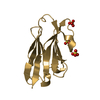
| Title | Crystal structure of an amyloid-beta binding single chain antibody G7 | ||||||
 Components Components | ANTIBODY G7 | ||||||
 Keywords Keywords | IMMUNE SYSTEM / ALZHEIMER'S DISEASE | ||||||
| Function / homology | Immunoglobulins / Immunoglobulin-like / Sandwich / Mainly Beta Function and homology information Function and homology information | ||||||
| Biological species |  | ||||||
| Method |  X-RAY DIFFRACTION / SYNCHROTRON / MOLECULAR REPLACEMENT / Resolution: 1.191 Å X-RAY DIFFRACTION / SYNCHROTRON / MOLECULAR REPLACEMENT / Resolution: 1.191 Å | ||||||
 Authors Authors | Beringer, D.X. / Dorresteijn, B. / Rutten, L. / Wienk, H. / el Khattabi, M. / Kroon-Batenburg, L.M.J. / Verrips, C.T. | ||||||
 Citation Citation | Journal: To be Published Title: Crystal Structure of an Amyloid-Beta Binding Single Chain Antibody G7 Authors: Beringer, D.X. / Dorresteijn, B. / Rutten, L. / Wienk, H. / El Khattabi, M. / Kroon-Batenburg, L.M.J. / Verrips, C.T. | ||||||
| History |
|
- Structure visualization
| Structure viewer | Molecule: MolmilJmol/JSmol |
|---|
- Downloads & links
-Download
| PDBx/mmCIF format | 4b41.cif.gz | 113.1 KB | Display | PDBx/mmCIF format |
|---|---|---|---|---|
| PDB format | pdb4b41.ent.gz | 88.3 KB | Display | PDB format |
| PDBx/mmJSON format | 4b41.json.gz | Tree view | PDBx/mmJSON format | |
| Others |  Other downloads Other downloads |
-Validation report
| Arichive directory | https://data.pdbj.org/pub/pdb/validation_reports/b4/4b41ftp://data.pdbj.org/pub/pdb/validation_reports/b4/4b41 | HTTPS FTP |
|---|
-Related structure data
| Related structure data | 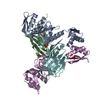 3ezjS S: Starting model for refinement |
|---|---|
| Similar structure data |


-Links
 PDBj
PDBj

- Assembly
| Deposited unit | 
| ||||||||
|---|---|---|---|---|---|---|---|---|---|
| 1 | 
| ||||||||
| 2 | 
| ||||||||
| Unit cell |
|
-Components
| #1: Antibody | Mass: 12667.176 Da / Num. of mol.: 2 Source method: isolated from a genetically manipulated source Source: (gene. exp.)  #2: Chemical | ChemComp-CL /   Mass: 35.453 Da / Num. of mol.: 4 / Source method: obtained synthetically / Formula: Cl Mass: 35.453 Da / Num. of mol.: 4 / Source method: obtained synthetically / Formula: Cl#3: Chemical | ChemComp-GOL / |   Mass: 92.094 Da / Num. of mol.: 1 / Source method: obtained synthetically / Formula: C3H8O3 Mass: 92.094 Da / Num. of mol.: 1 / Source method: obtained synthetically / Formula: C3H8O3#4: Water | ChemComp-HOH / |  Mass: 18.015 Da / Num. of mol.: 294 / Source method: isolated from a natural source / Formula: H2O Mass: 18.015 Da / Num. of mol.: 294 / Source method: isolated from a natural source / Formula: H2OHas protein modification | Y | |
|---|
-Experimental details
-Experiment
| Experiment | Method: X-RAY DIFFRACTION / Number of used crystals: 1 |
|---|
- Sample preparation
| Crystal | Density Matthews: 1.75 Å3/Da / Density % sol: 29.6 % / Description: NONE |
|---|---|
| Crystal grow | pH: 5.2 / Details: 0.1 M NA CITRATE PH 5.2, 0.2 NACL, 26-30% PEG8K |
-Data collection
| Diffraction | Mean temperature: 110 K |
|---|---|
| Diffraction source | Source: SYNCHROTRON / Site: SLS  / Beamline: X06SA / Wavelength: 1 / Beamline: X06SA / Wavelength: 1 |
| Detector | Type: DECTRIS PILATUS 6M / Detector: PIXEL / Date: Aug 19, 2010 / Details: DYNAMICALLY BENDABLE MIRROR |
| Radiation | Monochromator: LN2 COOLED FIXED-EXIT SI(111) MONOCHROMATOR / Protocol: SINGLE WAVELENGTH / Monochromatic (M) / Laue (L): M / Scattering type: x-ray |
| Radiation wavelength | Wavelength: 1 Å / Relative weight: 1 |
| Reflection | Resolution: 1.19→49.94 Å / Num. obs: 50156 / % possible obs: 85.1 % / Observed criterion σ(I): 6.7 / Redundancy: 3.4 % / Biso Wilson estimate: 10.26 Å2 / Rmerge(I) obs: 0.03 / Net I/σ(I): 18.6 |
| Reflection shell | Resolution: 1.19→1.26 Å / Redundancy: 3.3 % / Rmerge(I) obs: 0.15 / Mean I/σ(I) obs: 6.7 / % possible all: 62.6 |
- Processing
| Software |
| |||||||||||||||||||||||||||||||||||||||||||||||||||||||||||||||||||||||||||||||||||||||||||||||||||||||||||||||||||||||||||||||||||||
|---|---|---|---|---|---|---|---|---|---|---|---|---|---|---|---|---|---|---|---|---|---|---|---|---|---|---|---|---|---|---|---|---|---|---|---|---|---|---|---|---|---|---|---|---|---|---|---|---|---|---|---|---|---|---|---|---|---|---|---|---|---|---|---|---|---|---|---|---|---|---|---|---|---|---|---|---|---|---|---|---|---|---|---|---|---|---|---|---|---|---|---|---|---|---|---|---|---|---|---|---|---|---|---|---|---|---|---|---|---|---|---|---|---|---|---|---|---|---|---|---|---|---|---|---|---|---|---|---|---|---|---|---|---|---|
| Refinement | Method to determine structure: MOLECULAR REPLACEMENT Starting model: PDB ENTRY 3EZJ Resolution: 1.191→49.936 Å / SU ML: 0.09 / σ(F): 1.96 / Phase error: 18.48 / Stereochemistry target values: ML
| |||||||||||||||||||||||||||||||||||||||||||||||||||||||||||||||||||||||||||||||||||||||||||||||||||||||||||||||||||||||||||||||||||||
| Solvent computation | Shrinkage radii: 0.9 Å / VDW probe radii: 1.11 Å / Solvent model: FLAT BULK SOLVENT MODEL / Bsol: 0 Å2 / ksol: 0 e/Å3 | |||||||||||||||||||||||||||||||||||||||||||||||||||||||||||||||||||||||||||||||||||||||||||||||||||||||||||||||||||||||||||||||||||||
| Displacement parameters | Biso mean: 15.4 Å2 | |||||||||||||||||||||||||||||||||||||||||||||||||||||||||||||||||||||||||||||||||||||||||||||||||||||||||||||||||||||||||||||||||||||
| Refinement step | Cycle: LAST / Resolution: 1.191→49.936 Å
| |||||||||||||||||||||||||||||||||||||||||||||||||||||||||||||||||||||||||||||||||||||||||||||||||||||||||||||||||||||||||||||||||||||
| Refine LS restraints |
| |||||||||||||||||||||||||||||||||||||||||||||||||||||||||||||||||||||||||||||||||||||||||||||||||||||||||||||||||||||||||||||||||||||
| LS refinement shell |
|
