Mass: 18.015 Da / Num. of mol.: 208 / Source method: isolated from a natural source / Formula: D2O
-
Experimental details
-
Experiment
Experiment
Method
Number of used crystals
X-RAY DIFFRACTION
1
NEUTRON DIFFRACTION
-
Sample preparation
Crystal
Density Matthews: 2.16 Å3/Da / Density % sol: 43.07 % / Description: Thick rods.
Crystal grow
Temperature: 293 K / Method: vapor diffusion, sitting drop / pH: 8.5 Details: 1.2 M sodium citrate, 0.1 M Tris pH 8.5 in Hampton sandwich box set-ups. Drops were 150-250 uL and we set-up in a 1:1 ratio. To get the complex with SGC ligand we did "dry co-crystallization".
-
Data collection
Diffraction
ID
Mean temperature (K)
Crystal-ID
1
293
1
2
293
1
Diffraction source
Source
Site
Beamline
ID
Wavelength (Å)
SYNCHROTRON
ESRF
BM30A
1
1.04
NUCLEAR REACTOR
FRM II
BIODIFF
2
2.66
Detector
Type
ID
Detector
Date
ADSC QUANTUM 315r
1
CCD
Dec 15, 2016
MAATEL IMAGINE
2
IMAGE PLATE
Mar 1, 2018
Radiation
ID
Protocol
Monochromatic (M) / Laue (L)
Scattering type
Wavelength-ID
1
SINGLEWAVELENGTH
M
x-ray
1
2
SINGLEWAVELENGTH
M
neutron
2
Radiation wavelength
ID
Wavelength (Å)
Relative weight
1
1.04
1
2
2.66
1
Reflection
Entry-ID: 6GCY
Resolution (Å)
Num. obs
% possible obs (%)
Redundancy (%)
Rmerge(I) obs
Diffraction-ID
Net I/σ(I)
1.3-36
60056
97.6
7.3
0.051
1
22.3
2-50
16153
93
2.3
0.168
2
3.9
Reflection shell
Resolution (Å)
Rmerge(I) obs
Diffraction-ID
1.3-1.4
0.524
1
2-2.06
0.469
2
-
Processing
Software
Name
Version
Classification
PHENIX
(1.12_2829: ???)
refinement
HKL-2000
datareduction
HKL-2000
datascaling
PHENIX
phasing
Refinement
SU ML: 0.1 / Cross valid method: THROUGHOUT / Method to determine structure: MOLECULAR REPLACEMENT / Phase error: 19.66 / Shrinkage radii: 0.9 Å / VDW probe radii: 1.11 Å / Starting model: 4riu
In the structure databanks used in Yorodumi, some data are registered as the other names, "COVID-19 virus" and "2019-nCoV". Here are the details of the virus and the list of structure data.
Jan 31, 2019. EMDB accession codes are about to change! (news from PDBe EMDB page)
EMDB accession codes are about to change! (news from PDBe EMDB page)
The allocation of 4 digits for EMDB accession codes will soon come to an end. Whilst these codes will remain in use, new EMDB accession codes will include an additional digit and will expand incrementally as the available range of codes is exhausted. The current 4-digit format prefixed with “EMD-” (i.e. EMD-XXXX) will advance to a 5-digit format (i.e. EMD-XXXXX), and so on. It is currently estimated that the 4-digit codes will be depleted around Spring 2019, at which point the 5-digit format will come into force.
The EM Navigator/Yorodumi systems omit the EMD- prefix.
Related info.:Q: What is EMD? / ID/Accession-code notation in Yorodumi/EM Navigator
Yorodumi is a browser for structure data from EMDB, PDB, SASBDB, etc.
This page is also the successor to EM Navigator detail page, and also detail information page/front-end page for Omokage search.
The word "yorodu" (or yorozu) is an old Japanese word meaning "ten thousand". "mi" (miru) is to see.
Related info.:EMDB / PDB / SASBDB / Comparison of 3 databanks / Yorodumi Search / Aug 31, 2016. New EM Navigator & Yorodumi / Yorodumi Papers / Jmol/JSmol / Function and homology information / Changes in new EM Navigator and Yorodumi
 Movie
Movie Controller

 Yorodumi
 Open data
 Basic information
 Components
 Keywords
 Function and homology information
 Homo sapiens (human)
 X-RAY DIFFRACTION / NEUTRON DIFFRACTION /
  Authors
 Citation
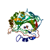
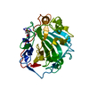
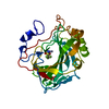
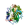
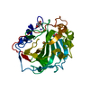
 Structure visualization
 Downloads & links
 Other downloads
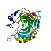


 PDBj
PDBj


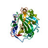
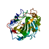
 Assembly
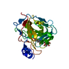


 Mass: 65.409 Da / Num. of mol.: 1 / Source method: obtained synthetically / Formula: Zn
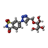
 Mass: 442.401 Da / Num. of mol.: 1 / Source method: obtained synthetically / Formula: C16H18N4O9S
 Mass: 18.015 Da / Num. of mol.: 208 / Source method: isolated from a natural source / Formula: D2O
 Sample preparation

 Processing