 Movie
Movie Controller

[English] 日本語
 Yorodumi
- PDB-6gcf: DNA binding domain of restriction endonuclease McrBC in complex w... -
+ Open data

- Basic information
| Entry | Database: PDB / ID: 6gcf | ||||||
|---|---|---|---|---|---|---|---|
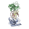
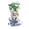
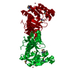
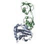
| Title | DNA binding domain of restriction endonuclease McrBC in complex with N4-methylcytosine DNA | ||||||
 Components Components |
| ||||||
 Keywords Keywords | DNA BINDING PROTEIN / N4-methylcytosine / restriction endonuclease / McrBC / base flipping | ||||||
| Function / homology |  Function and homology information Function and homology informationrestriction endodeoxyribonuclease activity / endonuclease complex / double-stranded methylated DNA binding / hemi-methylated DNA-binding / Hydrolases; Acting on ester bonds; Endodeoxyribonucleases producing 5'-phosphomonoesters / DNA restriction-modification system / DNA catabolic process / endonuclease activity / GTPase activity / GTP binding ...restriction endodeoxyribonuclease activity / endonuclease complex / double-stranded methylated DNA binding / hemi-methylated DNA-binding / Hydrolases; Acting on ester bonds; Endodeoxyribonucleases producing 5'-phosphomonoesters / DNA restriction-modification system / DNA catabolic process / endonuclease activity / GTPase activity / GTP binding / ATP hydrolysis activity / DNA binding / ATP binding / identical protein binding Similarity search - Function | ||||||
| Biological species |  synthetic construct (others) | ||||||
| Method |  X-RAY DIFFRACTION / SYNCHROTRON / MOLECULAR REPLACEMENT / Resolution: 1.55 Å X-RAY DIFFRACTION / SYNCHROTRON / MOLECULAR REPLACEMENT / Resolution: 1.55 Å | ||||||
 Authors Authors | Sasnauskas, G. | ||||||
| Funding support | Lithuania, 1items
| ||||||
 Citation Citation | Journal: FEBS Lett. / Year: 2018 Title: Recognition of modified cytosine variants by the DNA-binding domain of methyl-directed endonuclease McrBC. Authors: Zagorskaite, E. / Manakova, E. / Sasnauskas, G. | ||||||
| History |
|
- Structure visualization
| Structure viewer | Molecule: MolmilJmol/JSmol |
|---|
- Downloads & links
-Download
| PDBx/mmCIF format | 6gcf.cif.gz | 190.6 KB | Display | PDBx/mmCIF format |
|---|---|---|---|---|
| PDB format | pdb6gcf.ent.gz | 149.3 KB | Display | PDB format |
| PDBx/mmJSON format | 6gcf.json.gz | Tree view | PDBx/mmJSON format | |
| Others |  Other downloads Other downloads |
-Validation report
| Arichive directory | https://data.pdbj.org/pub/pdb/validation_reports/gc/6gcfftp://data.pdbj.org/pub/pdb/validation_reports/gc/6gcf | HTTPS FTP |
|---|
-Related structure data
| Related structure data |  6gcdC  6gceC  3sscS C: citing same article ( S: Starting model for refinement |
|---|---|
| Similar structure data |

-Links
 PDBj
PDBj


- Assembly
| Deposited unit | 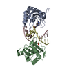
| ||||||||
|---|---|---|---|---|---|---|---|---|---|
| 1 |
| ||||||||
| Unit cell |
|
-Components
| #1: Protein | Mass: 19659.920 Da / Num. of mol.: 2 Source method: isolated from a genetically manipulated source Source: (gene. exp.) Production host: References: UniProt: P15005, Hydrolases; Acting on ester bonds; Endodeoxyribonucleases producing 5'-phosphomonoesters #2: DNA chain | | Mass: 3712.430 Da / Num. of mol.: 1 / Source method: obtained synthetically / Source: (synth.) synthetic construct (others) #3: DNA chain | | Mass: 3628.381 Da / Num. of mol.: 1 / Source method: obtained synthetically / Source: (synth.) synthetic construct (others) #4: Water | ChemComp-HOH / |  Mass: 18.015 Da / Num. of mol.: 264 / Source method: isolated from a natural source / Formula: H2O Mass: 18.015 Da / Num. of mol.: 264 / Source method: isolated from a natural source / Formula: H2O |
|---|
-Experimental details
-Experiment
| Experiment | Method: X-RAY DIFFRACTION / Number of used crystals: 1 |
|---|
- Sample preparation
| Crystal | Density Matthews: 1.96 Å3/Da / Density % sol: 37.39 % |
|---|---|
| Crystal grow | Temperature: 293 K / Method: vapor diffusion, sitting drop / pH: 7.5 / Details: 0.2 M LiCl, 17% PEG4000 |
-Data collection
| Diffraction | Mean temperature: 100 K |
|---|---|
| Diffraction source | Source: SYNCHROTRON / Site: PETRA III, EMBL c/o DESY  / Beamline: P13 (MX1) / Wavelength: 1.0332 Å / Beamline: P13 (MX1) / Wavelength: 1.0332 Å |
| Detector | Type: DECTRIS PILATUS3 6M / Detector: PIXEL / Date: Jun 26, 2015 |
| Radiation | Protocol: SINGLE WAVELENGTH / Monochromatic (M) / Laue (L): M / Scattering type: x-ray |
| Radiation wavelength | Wavelength: 1.0332 Å / Relative weight: 1 |
| Reflection | Resolution: 1.55→62.81 Å / Num. obs: 108317 / % possible obs: 99.88 % / Redundancy: 6.5 % / Biso Wilson estimate: 21.29 Å2 / Rmerge(I) obs: 0.04035 / Net I/σ(I): 28.36 |
| Reflection shell | Resolution: 1.55→1.605 Å / Redundancy: 6.2 % / Rmerge(I) obs: 0.4023 / Mean I/σ(I) obs: 5.38 / Num. unique obs: 10618 / % possible all: 99.16 |
- Processing
| Software |
| ||||||||||||||||||||||||||||||||||||||||||||||||||||||||||||||||||||||||||||||||||||||||||||||||||||||||||||||||||||||||||||||||||||||||||||||||||||||||||||||||||||||||||||||||||||||||||||||||||||
|---|---|---|---|---|---|---|---|---|---|---|---|---|---|---|---|---|---|---|---|---|---|---|---|---|---|---|---|---|---|---|---|---|---|---|---|---|---|---|---|---|---|---|---|---|---|---|---|---|---|---|---|---|---|---|---|---|---|---|---|---|---|---|---|---|---|---|---|---|---|---|---|---|---|---|---|---|---|---|---|---|---|---|---|---|---|---|---|---|---|---|---|---|---|---|---|---|---|---|---|---|---|---|---|---|---|---|---|---|---|---|---|---|---|---|---|---|---|---|---|---|---|---|---|---|---|---|---|---|---|---|---|---|---|---|---|---|---|---|---|---|---|---|---|---|---|---|---|---|---|---|---|---|---|---|---|---|---|---|---|---|---|---|---|---|---|---|---|---|---|---|---|---|---|---|---|---|---|---|---|---|---|---|---|---|---|---|---|---|---|---|---|---|---|---|---|---|---|
| Refinement | Method to determine structure: MOLECULAR REPLACEMENT Starting model: 3ssc Resolution: 1.55→62.81 Å / SU ML: 0.14 / Cross valid method: FREE R-VALUE / Phase error: 17.01
| ||||||||||||||||||||||||||||||||||||||||||||||||||||||||||||||||||||||||||||||||||||||||||||||||||||||||||||||||||||||||||||||||||||||||||||||||||||||||||||||||||||||||||||||||||||||||||||||||||||
| Solvent computation | Shrinkage radii: 0.9 Å / VDW probe radii: 1.11 Å | ||||||||||||||||||||||||||||||||||||||||||||||||||||||||||||||||||||||||||||||||||||||||||||||||||||||||||||||||||||||||||||||||||||||||||||||||||||||||||||||||||||||||||||||||||||||||||||||||||||
| Refinement step | Cycle: LAST / Resolution: 1.55→62.81 Å
| ||||||||||||||||||||||||||||||||||||||||||||||||||||||||||||||||||||||||||||||||||||||||||||||||||||||||||||||||||||||||||||||||||||||||||||||||||||||||||||||||||||||||||||||||||||||||||||||||||||
| Refine LS restraints |
| ||||||||||||||||||||||||||||||||||||||||||||||||||||||||||||||||||||||||||||||||||||||||||||||||||||||||||||||||||||||||||||||||||||||||||||||||||||||||||||||||||||||||||||||||||||||||||||||||||||
| LS refinement shell |
|
