 Movie
Movie Controller

[English] 日本語
 Yorodumi
- PDB-3zvm: The structural basis for substrate recognition by mammalian polyn... -
+ Open data

- Basic information
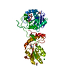
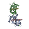
| Entry | Database: PDB / ID: 3zvm | ||||||
|---|---|---|---|---|---|---|---|
| Title | The structural basis for substrate recognition by mammalian polynucleotide kinase 3' phosphatase | ||||||
 Components Components |
| ||||||
 Keywords Keywords | HYDROLASE/TRANSFERASE/DNA / HYDROLASE-TRANSFERASE-DNA COMPLEX / BASE EXCISION REPAIR / BER / NON-HOMOLOGOUS END-JOINING / NHEJ / DNA REPAIR / CANCER | ||||||
| Function / homology |  Function and homology information Function and homology informationpolynucleotide 3'-phosphatase / polynucleotide 3'-phosphatase activity / polynucleotide 5'-hydroxyl-kinase / ATP-dependent polydeoxyribonucleotide 5'-hydroxyl-kinase activity / APEX1-Independent Resolution of AP Sites via the Single Nucleotide Replacement Pathway / positive regulation of double-strand break repair via nonhomologous end joining / SCF ubiquitin ligase complex / positive regulation of telomere maintenance / protein K63-linked ubiquitination / ubiquitin-like ligase-substrate adaptor activity ...polynucleotide 3'-phosphatase / polynucleotide 3'-phosphatase activity / polynucleotide 5'-hydroxyl-kinase / ATP-dependent polydeoxyribonucleotide 5'-hydroxyl-kinase activity / APEX1-Independent Resolution of AP Sites via the Single Nucleotide Replacement Pathway / positive regulation of double-strand break repair via nonhomologous end joining / SCF ubiquitin ligase complex / positive regulation of telomere maintenance / protein K63-linked ubiquitination / ubiquitin-like ligase-substrate adaptor activity / double-strand break repair via nonhomologous end joining / site of double-strand break / response to oxidative stress / DNA repair / DNA damage response / nucleolus / nucleoplasm / ATP binding / nucleus Similarity search - Function | ||||||
| Biological species |  synthetic construct (others) | ||||||
| Method |  X-RAY DIFFRACTION / MOLECULAR REPLACEMENT / Resolution: 1.997 Å X-RAY DIFFRACTION / MOLECULAR REPLACEMENT / Resolution: 1.997 Å | ||||||
 Authors Authors | Garces, F. / Pearl, L.H. / Oliver, A.W. | ||||||
 Citation Citation | Journal: Mol. Cell / Year: 2011 Title: The structural basis for substrate recognition by mammalian polynucleotide kinase 3' phosphatase. Authors: Garces, F. / Pearl, L.H. / Oliver, A.W. | ||||||
| History |
|
- Structure visualization
| Structure viewer | Molecule: MolmilJmol/JSmol |
|---|
- Downloads & links
-Download
| PDBx/mmCIF format | 3zvm.cif.gz | 202.2 KB | Display | PDBx/mmCIF format |
|---|---|---|---|---|
| PDB format | pdb3zvm.ent.gz | 155.7 KB | Display | PDB format |
| PDBx/mmJSON format | 3zvm.json.gz | Tree view | PDBx/mmJSON format | |
| Others |  Other downloads Other downloads |
-Validation report
| Arichive directory | https://data.pdbj.org/pub/pdb/validation_reports/zv/3zvmftp://data.pdbj.org/pub/pdb/validation_reports/zv/3zvm | HTTPS FTP |
|---|
-Related structure data
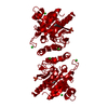
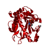
| Related structure data |  3zvlC  3zvnC  1yj5S C: citing same article ( S: Starting model for refinement |
|---|---|
| Similar structure data |

-Links
 PDBj
PDBj


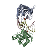
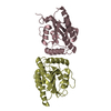
- Assembly
| Deposited unit | 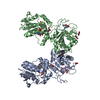
| ||||||||
|---|---|---|---|---|---|---|---|---|---|
| 1 | 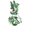
| ||||||||
| 2 | 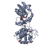
| ||||||||
| Unit cell |
|
-Components
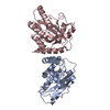
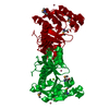
-Protein / DNA chain , 2 types, 4 molecules ABEF
| #1: Protein | Mass: 45932.227 Da / Num. of mol.: 2 Fragment: POLYNUCLEOTIDE 3'-PHOSPHATASE AND POLYNUCLEOTIDE 5'-HYDROXYL-KINASE DOMAINS, RESIDUES 111-522 Source method: isolated from a genetically manipulated source Source: (gene. exp.)  References: UniProt: Q9JLV6, polynucleotide 3'-phosphatase, polynucleotide 5'-hydroxyl-kinase #2: DNA chain | Mass: 1480.012 Da / Num. of mol.: 2 / Source method: obtained synthetically / Source: (synth.) synthetic construct (others) |
|---|
-Non-polymers , 6 types, 1214 molecules 


| #3: Chemical |  Mass: 427.201 Da / Num. of mol.: 2 / Source method: obtained synthetically / Formula: C10H15N5O10P2 / Comment: ADP, energy-carrying molecule*YM Mass: 427.201 Da / Num. of mol.: 2 / Source method: obtained synthetically / Formula: C10H15N5O10P2 / Comment: ADP, energy-carrying molecule*YM#4: Chemical |  Mass: 238.305 Da / Num. of mol.: 2 / Source method: obtained synthetically / Formula: C8H18N2O4S / Comment: pH buffer*YM Mass: 238.305 Da / Num. of mol.: 2 / Source method: obtained synthetically / Formula: C8H18N2O4S / Comment: pH buffer*YM#5: Chemical | ChemComp-MG /  Mass: 24.305 Da / Num. of mol.: 4 / Source method: obtained synthetically / Formula: Mg Mass: 24.305 Da / Num. of mol.: 4 / Source method: obtained synthetically / Formula: Mg#6: Chemical | ChemComp-GOL /  Mass: 92.094 Da / Num. of mol.: 8 / Source method: obtained synthetically / Formula: C3H8O3 Mass: 92.094 Da / Num. of mol.: 8 / Source method: obtained synthetically / Formula: C3H8O3#7: Chemical |  Mass: 59.044 Da / Num. of mol.: 2 / Source method: obtained synthetically / Formula: C2H3O2 Mass: 59.044 Da / Num. of mol.: 2 / Source method: obtained synthetically / Formula: C2H3O2#8: Water | ChemComp-HOH / | Mass: 18.015 Da / Num. of mol.: 1196 / Source method: isolated from a natural source / Formula: H2O |
|---|
-Experimental details
-Experiment
| Experiment | Method: X-RAY DIFFRACTION / Number of used crystals: 1 |
|---|
- Sample preparation
| Crystal | Density Matthews: 2.32 Å3/Da / Density % sol: 47 % / Description: NONE |
|---|---|
| Crystal grow | Details: 0.1 M HEPES PH 7.0, 18% (W/V) PEG 12000 |
-Data collection
| Diffraction | Mean temperature: 100 K |
|---|---|
| Diffraction source | Source: ROTATING ANODE / Type: BRUKER AXS MICROSTAR-H / Wavelength: 1.5418 |
| Detector | Type: BRUKER / Detector: CCD / Date: Feb 15, 2010 |
| Radiation | Protocol: SINGLE WAVELENGTH / Monochromatic (M) / Laue (L): M / Scattering type: x-ray |
| Radiation wavelength | Wavelength: 1.5418 Å / Relative weight: 1 |
| Reflection | Resolution: 2→38.5 Å / Num. obs: 60655 / % possible obs: 99.8 % / Observed criterion σ(I): 0 / Redundancy: 5.12 % / Biso Wilson estimate: 15.22 Å2 / Rmerge(I) obs: 0.12 / Net I/σ(I): 9.32 |
| Reflection shell | Resolution: 2→2.04 Å / Redundancy: 3.75 % / Rmerge(I) obs: 0.31 / Mean I/σ(I) obs: 3.5 / % possible all: 98.5 |
- Processing
| Software |
| |||||||||||||||||||||||||||||||||||||||||||||||||||||||||||||||||||||||||||||
|---|---|---|---|---|---|---|---|---|---|---|---|---|---|---|---|---|---|---|---|---|---|---|---|---|---|---|---|---|---|---|---|---|---|---|---|---|---|---|---|---|---|---|---|---|---|---|---|---|---|---|---|---|---|---|---|---|---|---|---|---|---|---|---|---|---|---|---|---|---|---|---|---|---|---|---|---|---|---|
| Refinement | Method to determine structure: MOLECULAR REPLACEMENT Starting model: PDB ENTRY 1YJ5 Resolution: 1.997→38.49 Å / SU ML: 0.23 / σ(F): 1.35 / Phase error: 17.98 / Stereochemistry target values: ML
| |||||||||||||||||||||||||||||||||||||||||||||||||||||||||||||||||||||||||||||
| Solvent computation | Shrinkage radii: 0.9 Å / VDW probe radii: 1.11 Å / Solvent model: FLAT BULK SOLVENT MODEL / Bsol: 43.72 Å2 / ksol: 0.346 e/Å3 | |||||||||||||||||||||||||||||||||||||||||||||||||||||||||||||||||||||||||||||
| Displacement parameters |
| |||||||||||||||||||||||||||||||||||||||||||||||||||||||||||||||||||||||||||||
| Refinement step | Cycle: LAST / Resolution: 1.997→38.49 Å
| |||||||||||||||||||||||||||||||||||||||||||||||||||||||||||||||||||||||||||||
| Refine LS restraints |
| |||||||||||||||||||||||||||||||||||||||||||||||||||||||||||||||||||||||||||||
| LS refinement shell |
|
