 Movie
Movie Controller

[English] 日本語
 Yorodumi
- PDB-6g7x: Crystal structure of H. pylori purine nucleoside phosphorylase so... -
+ Open data

- Basic information
| Entry | Database: PDB / ID: 6g7x | ||||||
|---|---|---|---|---|---|---|---|
| Title | Crystal structure of H. pylori purine nucleoside phosphorylase soaked in PO4 | ||||||
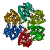
 Components Components | Purine nucleoside phosphorylase DeoD-type | ||||||
 Keywords Keywords | TRANSFERASE / Purine nucleoside phosphorylase / H. pylori | ||||||
| Function / homology |  Function and homology information Function and homology informationpurine-nucleoside phosphorylase / purine-nucleoside phosphorylase activity / purine nucleoside catabolic process / cytosol Similarity search - Function | ||||||
| Biological species |   Helicobacter pylori (bacteria) Helicobacter pylori (bacteria) | ||||||
| Method |  X-RAY DIFFRACTION / SYNCHROTRON / MOLECULAR REPLACEMENT / Resolution: 1.762 Å X-RAY DIFFRACTION / SYNCHROTRON / MOLECULAR REPLACEMENT / Resolution: 1.762 Å | ||||||
 Authors Authors | Stefanic, Z. | ||||||
| Funding support |  Croatia, 1items Croatia, 1items
| ||||||
 Citation Citation | Journal: Croatica Chemica Acta / Year: 2018 Title: The Role of Phosphate Binding in Purine Nucleoside Phosphorylase of Helicobacter pylori Authors: Bosnjakovic, M. / Lescic Asler, I. / Stefanic, Z. | ||||||
| History |
|
- Structure visualization
| Structure viewer | Molecule: MolmilJmol/JSmol |
|---|
- Downloads & links
-Download
| PDBx/mmCIF format | 6g7x.cif.gz | 298.9 KB | Display | PDBx/mmCIF format |
|---|---|---|---|---|
| PDB format | pdb6g7x.ent.gz | 242.1 KB | Display | PDB format |
| PDBx/mmJSON format | 6g7x.json.gz | Tree view | PDBx/mmJSON format | |
| Others |  Other downloads Other downloads |
-Validation report
| Arichive directory | https://data.pdbj.org/pub/pdb/validation_reports/g7/6g7xftp://data.pdbj.org/pub/pdb/validation_reports/g7/6g7x | HTTPS FTP |
|---|
-Related structure data
| Related structure data |  6f52S S: Starting model for refinement |
|---|---|
| Similar structure data |

-Links
 PDBj
PDBj


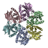
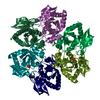
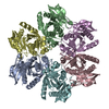
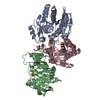
- Assembly
| Deposited unit | 
| ||||||||
|---|---|---|---|---|---|---|---|---|---|
| 1 |
| ||||||||
| Unit cell |
|
-Components
| #1: Protein | Mass: 25818.105 Da / Num. of mol.: 6 Source method: isolated from a genetically manipulated source Source: (gene. exp.) Helicobacter pylori (strain ATCC 700392 / 26695) (bacteria)Gene: deoD, HP_1178 / Plasmid: pET21b / Production host: References: UniProt: P56463, purine-nucleoside phosphorylase #2: Chemical | ChemComp-IMD /   Mass: 69.085 Da / Num. of mol.: 23 / Source method: obtained synthetically / Formula: C3H5N2 Mass: 69.085 Da / Num. of mol.: 23 / Source method: obtained synthetically / Formula: C3H5N2#3: Chemical | ChemComp-PO4 /   Mass: 94.971 Da / Num. of mol.: 6 / Source method: obtained synthetically / Formula: PO4 Mass: 94.971 Da / Num. of mol.: 6 / Source method: obtained synthetically / Formula: PO4#4: Water | ChemComp-HOH / |  Mass: 18.015 Da / Num. of mol.: 1027 / Source method: isolated from a natural source / Formula: H2O Mass: 18.015 Da / Num. of mol.: 1027 / Source method: isolated from a natural source / Formula: H2O |
|---|
-Experimental details
-Experiment
| Experiment | Method: X-RAY DIFFRACTION / Number of used crystals: 1 |
|---|
- Sample preparation
| Crystal | Density Matthews: 2.23 Å3/Da / Density % sol: 44.88 % |
|---|---|
| Crystal grow | Temperature: 291 K / Method: vapor diffusion, hanging drop / pH: 7 Details: 0.2 mol L-1 imidazole, 40% (w/v) polypropylene glycol (PPG) 400 |
-Data collection
| Diffraction | Mean temperature: 100 K |
|---|---|
| Diffraction source | Source: SYNCHROTRON / Site: ELETTRA  / Beamline: 5.2R / Wavelength: 1 Å / Beamline: 5.2R / Wavelength: 1 Å |
| Detector | Type: DECTRIS PILATUS 2M / Detector: PIXEL / Date: Jul 11, 2017 |
| Radiation | Protocol: SINGLE WAVELENGTH / Monochromatic (M) / Laue (L): M / Scattering type: x-ray |
| Radiation wavelength | Wavelength: 1 Å / Relative weight: 1 |
| Reflection | Resolution: 1.76→60.64 Å / Num. obs: 133830 / % possible obs: 99.7 % / Redundancy: 6.2 % / CC1/2: 0.997 / Rmerge(I) obs: 0.093 / Net I/σ(I): 13 |
| Reflection shell | Resolution: 1.76→1.86 Å / Redundancy: 5.7 % / Rmerge(I) obs: 0.348 / Num. unique obs: 19209 / CC1/2: 0.936 / % possible all: 98.3 |
- Processing
| Software |
| |||||||||||||||||||||||||||||||||||||||||||||||||||||||||||||||||||||||||||||||||||||||||||||||||||||||||
|---|---|---|---|---|---|---|---|---|---|---|---|---|---|---|---|---|---|---|---|---|---|---|---|---|---|---|---|---|---|---|---|---|---|---|---|---|---|---|---|---|---|---|---|---|---|---|---|---|---|---|---|---|---|---|---|---|---|---|---|---|---|---|---|---|---|---|---|---|---|---|---|---|---|---|---|---|---|---|---|---|---|---|---|---|---|---|---|---|---|---|---|---|---|---|---|---|---|---|---|---|---|---|---|---|---|---|
| Refinement | Method to determine structure: MOLECULAR REPLACEMENT Starting model: 6F52 Resolution: 1.762→46.753 Å / SU ML: 0.18 / Cross valid method: FREE R-VALUE / σ(F): 1.35 / Phase error: 19.38
| |||||||||||||||||||||||||||||||||||||||||||||||||||||||||||||||||||||||||||||||||||||||||||||||||||||||||
| Solvent computation | Shrinkage radii: 0.9 Å / VDW probe radii: 1.11 Å | |||||||||||||||||||||||||||||||||||||||||||||||||||||||||||||||||||||||||||||||||||||||||||||||||||||||||
| Displacement parameters | Biso max: 72.07 Å2 / Biso mean: 18.9557 Å2 / Biso min: 5.6 Å2 | |||||||||||||||||||||||||||||||||||||||||||||||||||||||||||||||||||||||||||||||||||||||||||||||||||||||||
| Refinement step | Cycle: final / Resolution: 1.762→46.753 Å
| |||||||||||||||||||||||||||||||||||||||||||||||||||||||||||||||||||||||||||||||||||||||||||||||||||||||||
| Refine LS restraints |
| |||||||||||||||||||||||||||||||||||||||||||||||||||||||||||||||||||||||||||||||||||||||||||||||||||||||||
| LS refinement shell | Refine-ID: X-RAY DIFFRACTION / Total num. of bins used: 14
|
