Protocol: SINGLE WAVELENGTH / Monochromatic (M) / Laue (L): M / Scattering type: x-ray
Radiation wavelength
Wavelength: 0.9791 Å / Relative weight: 1
Reflection
Redundancy: 3.9 % / Number: 126748 / Rmerge(I) obs: 0.133 / Χ2: 1.05 / D res high: 2.2 Å / D res low: 50 Å / Num. obs: 32265 / % possible obs: 88.8
Diffraction reflection shell
Highest resolution (Å)
Lowest resolution (Å)
ID
Rmerge(I) obs
Chi squared
Redundancy
5.97
50
1
0.065
0.754
4.5
4.74
5.97
1
0.088
1.086
4.5
4.14
4.74
1
0.083
0.962
4.7
3.76
4.14
1
0.096
1.015
4.8
3.49
3.76
1
0.116
1.047
4.8
3.29
3.49
1
0.14
1.081
4.8
3.12
3.29
1
0.175
1.111
4.8
2.99
3.12
1
0.22
1.102
4.6
2.87
2.99
1
0.283
1.107
4.5
2.77
2.87
1
0.397
1.064
4.4
2.69
2.77
1
0.514
1.084
4.3
2.61
2.69
1
0.55
1.087
4.1
2.54
2.61
1
0.621
1.066
3.8
2.48
2.54
1
0.719
1.087
3.1
2.42
2.48
1
0.62
1.042
2.5
2.37
2.42
1
0.703
1.103
2.3
2.32
2.37
1
0.753
1.089
2.2
2.28
2.32
1
0.763
1.061
1.9
2.24
2.28
1
0.655
1.126
1.9
2.2
2.24
1
0.948
1.087
1.8
Reflection
Resolution: 2.2→50 Å / Num. obs: 32265 / % possible obs: 88.8 % / Redundancy: 3.9 % / Biso Wilson estimate: 38.84 Å2 / Rmerge(I) obs: 0.133 / Χ2: 1.048 / Net I/av σ(I): 8.954 / Net I/σ(I): 7.3 / Num. measured all: 126748
Reflection shell
Diffraction-ID: 1 / Rejects: _
Resolution (Å)
Redundancy (%)
Rmerge(I) obs
Num. unique all
Χ2
% possible all
2.2-2.24
1.8
0.948
803
1.087
43.9
2.24-2.28
1.9
0.655
936
1.126
52
2.28-2.32
1.9
0.763
1094
1.061
59.9
2.32-2.37
2.2
0.753
1163
1.089
63.8
2.37-2.42
2.3
0.703
1410
1.103
78.7
2.42-2.48
2.5
0.62
1583
1.042
86.8
2.48-2.54
3.1
0.719
1772
1.087
97.8
2.54-2.61
3.8
0.621
1831
1.066
99.6
2.61-2.69
4.1
0.55
1790
1.087
99.7
2.69-2.77
4.3
0.514
1788
1.084
99.4
2.77-2.87
4.4
0.397
1803
1.064
99.4
2.87-2.99
4.5
0.283
1819
1.107
99.7
2.99-3.12
4.6
0.22
1806
1.102
99.8
3.12-3.29
4.8
0.175
1822
1.111
99.9
3.29-3.49
4.8
0.14
1822
1.081
99.9
3.49-3.76
4.8
0.116
1820
1.047
99.9
3.76-4.14
4.8
0.096
1794
1.015
99.9
4.14-4.74
4.7
0.083
1840
0.962
99.9
4.74-5.97
4.5
0.088
1788
1.086
99.6
5.97-50
4.5
0.065
1781
0.754
96.8
-
Phasing
Phasing
Method: SAD
-
Processing
Software
Name
Version
Classification
SCALEPACK
datascaling
SHELX
phasing
RESOLVE
phasing
PDB_EXTRACT
3.14
dataextraction
Coot
modelbuilding
PHENIX
(phenix.refine: 1.9_1692)
refinement
SHELXD
phasing
Refinement
Method to determine structure: SAD / Resolution: 2.399→33.895 Å / SU ML: 0.21 / Cross valid method: THROUGHOUT / σ(F): 0.01 / Phase error: 23.94 / Stereochemistry target values: ML
Rfactor
Num. reflection
% reflection
Rfree
0.1926
1311
4.72 %
Rwork
0.1664
26475
-
obs
0.1677
27786
99.3 %
Solvent computation
Shrinkage radii: 0.9 Å / VDW probe radii: 1.11 Å / Solvent model: FLAT BULK SOLVENT MODEL
In the structure databanks used in Yorodumi, some data are registered as the other names, "COVID-19 virus" and "2019-nCoV". Here are the details of the virus and the list of structure data.
Jan 31, 2019. EMDB accession codes are about to change! (news from PDBe EMDB page)
EMDB accession codes are about to change! (news from PDBe EMDB page)
The allocation of 4 digits for EMDB accession codes will soon come to an end. Whilst these codes will remain in use, new EMDB accession codes will include an additional digit and will expand incrementally as the available range of codes is exhausted. The current 4-digit format prefixed with “EMD-” (i.e. EMD-XXXX) will advance to a 5-digit format (i.e. EMD-XXXXX), and so on. It is currently estimated that the 4-digit codes will be depleted around Spring 2019, at which point the 5-digit format will come into force.
The EM Navigator/Yorodumi systems omit the EMD- prefix.
Related info.:Q: What is EMD? / ID/Accession-code notation in Yorodumi/EM Navigator
Yorodumi is a browser for structure data from EMDB, PDB, SASBDB, etc.
This page is also the successor to EM Navigator detail page, and also detail information page/front-end page for Omokage search.
The word "yorodu" (or yorozu) is an old Japanese word meaning "ten thousand". "mi" (miru) is to see.
Related info.:EMDB / PDB / SASBDB / Comparison of 3 databanks / Yorodumi Search / Aug 31, 2016. New EM Navigator & Yorodumi / Yorodumi Papers / Jmol/JSmol / Function and homology information / Changes in new EM Navigator and Yorodumi
 Movie
Movie Controller

 Yorodumi
 Open data
 Basic information
 Components
 Keywords
 Function and homology information
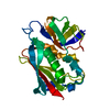
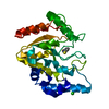
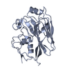
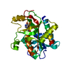
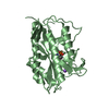
 Streptococcus agalactiae LMG 15084 (bacteria)
 X-RAY DIFFRACTION /
  Authors
 Citation
 Structure visualization
 Downloads & links
 Other downloads
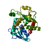


 PDBj
PDBj Assembly
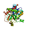

 Mass: 96.063 Da / Num. of mol.: 3 / Source method: obtained synthetically / Formula: SO4
 Mass: 18.015 Da / Num. of mol.: 91 / Source method: isolated from a natural source / Formula: H2O
 Sample preparation
 / Beamline: 31-ID / Wavelength: 0.9791 Å
 Processing