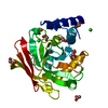
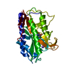
base-excision repair, AP site formation via deaminated base removal / uracil-DNA glycosylase / uracil DNA N-glycosylase activity / cytoplasm Similarity search - Function
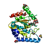
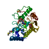
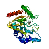
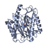
Uracil-DNA glycosylase family 1 / Uracil DNA glycosylase superfamily / UreE urease accessory protein, C-terminal domain / Uracil-DNA Glycosylase, subunit E / Uracil-DNA glycosylase-like domain / Uracil-DNA glycosylase, active site / Uracil-DNA glycosylase signature. / Uracil-DNA glycosylase-like / Uracil DNA glycosylase superfamily / Uracil-DNA glycosylase-like domain superfamily ...Uracil-DNA glycosylase family 1 / Uracil DNA glycosylase superfamily / UreE urease accessory protein, C-terminal domain / Uracil-DNA Glycosylase, subunit E / Uracil-DNA glycosylase-like domain / Uracil-DNA glycosylase, active site / Uracil-DNA glycosylase signature. / Uracil-DNA glycosylase-like / Uracil DNA glycosylase superfamily / Uracil-DNA glycosylase-like domain superfamily / 3-Layer(aba) Sandwich / Alpha Beta Similarity search - Domain/homology
Resolution: 2.95→48.73 Å / Cor.coef. Fo:Fc: 0.89 / Cor.coef. Fo:Fc free: 0.84 / SU B: 24.305 / SU ML: 0.479 / Cross valid method: THROUGHOUT / ESU R Free: 0.519 / Stereochemistry target values: MAXIMUM LIKELIHOOD / Details: HYDROGENS HAVE BEEN ADDED IN THE RIDING POSITIONS.
Rfactor
Num. reflection
% reflection
Selection details
Rfree
0.29516
252
4.5 %
RANDOM
Rwork
0.23779
-
-
-
obs
0.24045
5313
99.3 %
-
Solvent computation
Ion probe radii: 0.8 Å / Shrinkage radii: 0.8 Å / VDW probe radii: 1.2 Å / Solvent model: MASK
 Movie
Movie Controller


 Open data
 Basic information
 Components
 Keywords
 Function and homology information
 X-RAY DIFFRACTION /
  Authors
 Citation
 Structure visualization
 Downloads & links
 Other downloads


 PDBj
PDBj Assembly
 Sample preparation
 / Beamline: ID29 / Wavelength: 0.9791
  Processing