 Movie
Movie Controller

[English] 日本語
 Yorodumi
- PDB-4da0: Crystal structure of the hexameric purine nucleoside phosphorylas... -
+ Open data

- Basic information
| Entry | Database: PDB / ID: 4da0 | ||||||
|---|---|---|---|---|---|---|---|
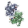
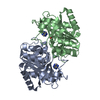
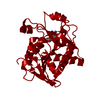
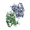
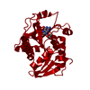
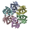
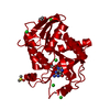
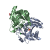
| Title | Crystal structure of the hexameric purine nucleoside phosphorylase from Bacillus subtilis in complex with 2'-deoxyguanosine | ||||||
 Components Components | Purine nucleoside phosphorylase deoD-type | ||||||
 Keywords Keywords | TRANSFERASE / Phosphorylase/hydrolase-like | ||||||
| Function / homology |  Function and homology information Function and homology informationpurine-nucleoside phosphorylase / purine-nucleoside phosphorylase activity / purine nucleoside catabolic process / cytosol Similarity search - Function | ||||||
| Biological species |  | ||||||
| Method |  X-RAY DIFFRACTION / SYNCHROTRON / MOLECULAR REPLACEMENT / Resolution: 2.95 Å X-RAY DIFFRACTION / SYNCHROTRON / MOLECULAR REPLACEMENT / Resolution: 2.95 Å | ||||||
 Authors Authors | Martins, N.H. / Giuseppe, P.O. / Meza, A.N. / Murakami, M.T. | ||||||
 Citation Citation | Journal: Plos One / Year: 2012 Title: Insights into phosphate cooperativity and influence of substrate modifications on binding and catalysis of hexameric purine nucleoside phosphorylases. Authors: de Giuseppe, P.O. / Martins, N.H. / Meza, A.N. / Dos Santos, C.R. / Pereira, H.D. / Murakami, M.T. | ||||||
| History |
|
- Structure visualization
| Structure viewer | Molecule: MolmilJmol/JSmol |
|---|
- Downloads & links
-Download
| PDBx/mmCIF format | 4da0.cif.gz | 57.9 KB | Display | PDBx/mmCIF format |
|---|---|---|---|---|
| PDB format | pdb4da0.ent.gz | 41.6 KB | Display | PDB format |
| PDBx/mmJSON format | 4da0.json.gz | Tree view | PDBx/mmJSON format | |
| Others |  Other downloads Other downloads |
-Validation report
| Arichive directory | https://data.pdbj.org/pub/pdb/validation_reports/da/4da0ftp://data.pdbj.org/pub/pdb/validation_reports/da/4da0 | HTTPS FTP |
|---|
-Related structure data
| Related structure data |  4d8vC  4d8xC  4d8yC  4d98C  4d9hC  4da6C  4da7C  4da8C  4dabC  4daeC  4danC  4daoC  4darC C: citing same article ( |
|---|---|
| Similar structure data |


-Links
 PDBj
PDBj- Assembly
| Deposited unit | 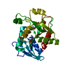
| ||||||||
|---|---|---|---|---|---|---|---|---|---|
| 1 | x 6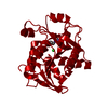
| ||||||||
| Unit cell |
|
-Components
| #1: Protein | Mass: 27562.137 Da / Num. of mol.: 1 Source method: isolated from a genetically manipulated source Source: (gene. exp.) References: UniProt: O34925, purine-nucleoside phosphorylase |
|---|---|
| #2: Chemical | ChemComp-GNG /   Mass: 267.241 Da / Num. of mol.: 1 / Source method: obtained synthetically / Formula: C10H13N5O4 Mass: 267.241 Da / Num. of mol.: 1 / Source method: obtained synthetically / Formula: C10H13N5O4 |
| #3: Chemical | ChemComp-CL /   Mass: 35.453 Da / Num. of mol.: 1 / Source method: obtained synthetically / Formula: Cl Mass: 35.453 Da / Num. of mol.: 1 / Source method: obtained synthetically / Formula: Cl |
| Sequence details | AUTHORS STATE THAT THIS IS A CLONING ARTIFACT |
-Experimental details
-Experiment
| Experiment | Method: X-RAY DIFFRACTION / Number of used crystals: 1 |
|---|
- Sample preparation
| Crystal | Density Matthews: 2.72 Å3/Da / Density % sol: 54.86 % |
|---|---|
| Crystal grow | Temperature: 291 K / Method: vapor diffusion / pH: 4.6 Details: 0.1 M sodium acetate, 3.2 M sodium chloride, 5%(v/v) glycerol, pH 4.6, VAPOR DIFFUSION, temperature 291K |
-Data collection
| Diffraction | Mean temperature: 100 K | |||||||||||||||||||||||||||||||||||||||||||||||||||||||||||||||||||||||||||||
|---|---|---|---|---|---|---|---|---|---|---|---|---|---|---|---|---|---|---|---|---|---|---|---|---|---|---|---|---|---|---|---|---|---|---|---|---|---|---|---|---|---|---|---|---|---|---|---|---|---|---|---|---|---|---|---|---|---|---|---|---|---|---|---|---|---|---|---|---|---|---|---|---|---|---|---|---|---|---|
| Diffraction source | Source: SYNCHROTRON / Site: LNLS  / Beamline: W01B-MX2 / Beamline: W01B-MX2 | |||||||||||||||||||||||||||||||||||||||||||||||||||||||||||||||||||||||||||||
| Detector | Type: MARMOSAIC 225 mm CCD / Detector: CCD | |||||||||||||||||||||||||||||||||||||||||||||||||||||||||||||||||||||||||||||
| Radiation | Protocol: SINGLE WAVELENGTH / Monochromatic (M) / Laue (L): M / Scattering type: x-ray | |||||||||||||||||||||||||||||||||||||||||||||||||||||||||||||||||||||||||||||
| Radiation wavelength | Relative weight: 1 | |||||||||||||||||||||||||||||||||||||||||||||||||||||||||||||||||||||||||||||
| Reflection | Resolution: 2.95→50 Å / Num. obs: 6857 / % possible obs: 99.7 % / Redundancy: 10 % / Rmerge(I) obs: 0.127 / Χ2: 1.027 / Net I/σ(I): 7.8 | |||||||||||||||||||||||||||||||||||||||||||||||||||||||||||||||||||||||||||||
| Reflection shell |
|
- Processing
| Software |
| |||||||||||||||||||||||||||||||||||||||||||||||||||||||||||||||||
|---|---|---|---|---|---|---|---|---|---|---|---|---|---|---|---|---|---|---|---|---|---|---|---|---|---|---|---|---|---|---|---|---|---|---|---|---|---|---|---|---|---|---|---|---|---|---|---|---|---|---|---|---|---|---|---|---|---|---|---|---|---|---|---|---|---|---|
| Refinement | Method to determine structure: MOLECULAR REPLACEMENT / Resolution: 2.95→44.3 Å / Cor.coef. Fo:Fc: 0.919 / Cor.coef. Fo:Fc free: 0.867 / WRfactor Rfree: 0.3156 / WRfactor Rwork: 0.2242 / Occupancy max: 1 / Occupancy min: 1 / FOM work R set: 0.7742 / SU B: 21.279 / SU ML: 0.397 / SU Rfree: 0.4879 / Cross valid method: THROUGHOUT / σ(F): 0 / ESU R Free: 0.488 / Stereochemistry target values: MAXIMUM LIKELIHOOD / Details: HYDROGENS HAVE BEEN ADDED IN THE RIDING POSITIONS
| |||||||||||||||||||||||||||||||||||||||||||||||||||||||||||||||||
| Solvent computation | Ion probe radii: 0.8 Å / Shrinkage radii: 0.8 Å / VDW probe radii: 1.4 Å / Solvent model: MASK | |||||||||||||||||||||||||||||||||||||||||||||||||||||||||||||||||
| Displacement parameters | Biso max: 95.17 Å2 / Biso mean: 62.8993 Å2 / Biso min: 39.38 Å2
| |||||||||||||||||||||||||||||||||||||||||||||||||||||||||||||||||
| Refinement step | Cycle: LAST / Resolution: 2.95→44.3 Å
| |||||||||||||||||||||||||||||||||||||||||||||||||||||||||||||||||
| Refine LS restraints |
| |||||||||||||||||||||||||||||||||||||||||||||||||||||||||||||||||
| LS refinement shell | Resolution: 2.95→3.024 Å / Total num. of bins used: 20
|
