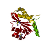
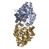
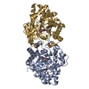
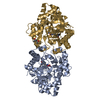
[English] 日本語
Yorodumi
- PDB-6fu6: Phosphotriesterase PTE_C23_2 -

+
Open data


ID or keywords:

Loading...

-
Basic information

Entry
Database: PDB / ID: 6fu6
TitlePhosphotriesterase PTE_C23_2
ComponentsParathion hydrolase
KeywordsHYDROLASE / Metalloenzyme / TIM barrel
Function / homology
Function and homology information


aryldialkylphosphatase / aryldialkylphosphatase activity / catabolic process / zinc ion binding / plasma membrane
Similarity search - Function
Aryldialkylphosphatase, zinc-binding site / Phosphotriesterase family signature 1. / Phosphotriesterase / Phosphotriesterase family / Phosphotriesterase family profile. / Metal-dependent hydrolases / Metal-dependent hydrolase / Twin arginine translocation (Tat) signal profile. / Twin-arginine translocation pathway, signal sequence / TIM Barrel ...Aryldialkylphosphatase, zinc-binding site / Phosphotriesterase family signature 1. / Phosphotriesterase / Phosphotriesterase family / Phosphotriesterase family profile. / Metal-dependent hydrolases / Metal-dependent hydrolase / Twin arginine translocation (Tat) signal profile. / Twin-arginine translocation pathway, signal sequence / TIM Barrel / Alpha-Beta Barrel / Alpha Beta
Similarity search - Domain/homology
FORMIC ACID / POLYACRYLIC ACID / Parathion hydrolase
Similarity search - Component
Biological speciesBrevundimonas diminuta (bacteria)
MethodX-RAY DIFFRACTION / MOLECULAR REPLACEMENT / Resolution: 1.95 Å
AuthorsDym, O. / Aggarwal, N. / Albeck, S. / Unger, T. / Hamer Rogotner, S. / Silman, I. / Leader, H. / Ashani, Y. / Goldsmith, M. / Greisen, P. ...Dym, O. / Aggarwal, N. / Albeck, S. / Unger, T. / Hamer Rogotner, S. / Silman, I. / Leader, H. / Ashani, Y. / Goldsmith, M. / Greisen, P. / Tawfik, D. / Sussman, L.J.
Funding support United States, 2items
OrganizationGrant numberCountry
DTRAHDTRA1-11-C-0026). United States
DTRACB10265 / HDTRA1724528 United States
CitationJournal: To Be Published
Title: Phosphotriesterase PTE_A53_4
Authors: Dym, O. / Aggarwal, N. / Albeck, S. / Unger, T. / Hamer Rogotner, S. / Silman, I. / Leader, H. / Ashani, Y. / Goldsmith, M. / Greisen, P. / Tawfik, D. / Sussman, L.J.
History
DepositionFeb 26, 2018Deposition site: PDBE / Processing site: PDBE
Revision 1.0Mar 20, 2019Provider: repository / Type: Initial release
Revision 1.1Jan 17, 2024Group: Data collection / Database references ...Data collection / Database references / Derived calculations / Refinement description
Category: chem_comp_atom / chem_comp_bond ...chem_comp_atom / chem_comp_bond / database_2 / pdbx_initial_refinement_model / struct_conn / struct_conn_type
Item: _database_2.pdbx_DOI / _database_2.pdbx_database_accession ..._database_2.pdbx_DOI / _database_2.pdbx_database_accession / _struct_conn.conn_type_id / _struct_conn.id / _struct_conn.pdbx_dist_value / _struct_conn.pdbx_leaving_atom_flag / _struct_conn.ptnr1_auth_asym_id / _struct_conn.ptnr1_auth_comp_id / _struct_conn.ptnr1_auth_seq_id / _struct_conn.ptnr1_label_asym_id / _struct_conn.ptnr1_label_atom_id / _struct_conn.ptnr1_label_comp_id / _struct_conn.ptnr1_label_seq_id / _struct_conn.ptnr2_auth_asym_id / _struct_conn.ptnr2_auth_comp_id / _struct_conn.ptnr2_auth_seq_id / _struct_conn.ptnr2_label_asym_id / _struct_conn.ptnr2_label_atom_id / _struct_conn.ptnr2_label_comp_id / _struct_conn_type.id

-
Structure visualization

Structure viewerMolecule:
MolmilJmol/JSmol

Downloads & links

-
Assembly

Deposited unit
A: Parathion hydrolase
B: Parathion hydrolase
hetero molecules


Theoretical massNumber of molelcules
Total (without water)72,61311
Polymers71,6132
Non-polymers1,0009
Water5,224290
1


  • Idetical with deposited unit
  • defined by author&software
  • Evidence: gel filtration
TypeNameSymmetry operationNumber
identity operation1_555x,y,z1
Buried area2990 Å2
ΔGint-88 kcal/mol
Surface area22990 Å2
MethodPISA
Unit cell
Length a, b, c (Å)54.660, 81.160, 70.660
Angle α, β, γ (deg.)90.00, 94.83, 90.00
Int Tables number4
Space group name H-MP1211

-
Components

#1: Protein Parathion hydrolase / Phosphotriesterase / PTE


Mass: 35806.605 Da / Num. of mol.: 2
Source method: isolated from a genetically manipulated source
Source: (gene. exp.) Brevundimonas diminuta (bacteria) / Gene: opd / Production host: Escherichia coli (E. coli) / References: UniProt: P0A434, aryldialkylphosphatase
#2: Chemical ChemComp-FMT / FORMIC ACID


Mass: 46.025 Da / Num. of mol.: 2 / Source method: obtained synthetically / Formula: CH2O2
#3: Chemical
ChemComp-ZN / ZINC ION


Mass: 65.409 Da / Num. of mol.: 5 / Source method: obtained synthetically / Formula: Zn
#4: Chemical ChemComp-TA8 / POLYACRYLIC ACID / OCTANE-1,3,5,7-TETRACARBOXYLIC ACID


Mass: 290.267 Da / Num. of mol.: 2 / Source method: obtained synthetically / Formula: C12H18O8
#5: Water ChemComp-HOH / water


Mass: 18.015 Da / Num. of mol.: 290 / Source method: isolated from a natural source / Formula: H2O

-
Experimental details

-
Experiment

ExperimentMethod: X-RAY DIFFRACTION / Number of used crystals: 1

-
Sample preparation

CrystalDensity Matthews: 2.18 Å3/Da / Density % sol: 43.6 %
Crystal growTemperature: 293 K / Method: vapor diffusion, hanging drop
Details: 0.05M HEPES pH=7.5 13% Polyacrylic acid 5100 Na 0.01M MgCl2

-
Data collection

DiffractionMean temperature: 100 K
Diffraction sourceSource: ROTATING ANODE / Type: RIGAKU RUH3R / Wavelength: 1.5415 Å
DetectorType: RIGAKU RAXIS IV++ / Detector: IMAGE PLATE / Date: May 25, 2014
RadiationProtocol: SINGLE WAVELENGTH / Monochromatic (M) / Laue (L): M / Scattering type: x-ray
Radiation wavelengthWavelength: 1.5415 Å / Relative weight: 1
ReflectionResolution: 1.95→70.41 Å / Num. obs: 44881 / % possible obs: 99.96 % / Observed criterion σ(F): 0 / Redundancy: 6.1 % / Biso Wilson estimate: 18.32 Å2 / Rmerge(I) obs: 0.05192 / Net I/σ(I): 11.84
Reflection shellResolution: 1.95→2.02 Å / Rmerge(I) obs: 0.248 / Mean I/σ(I) obs: 3.18 / Num. unique obs: 4458 / % possible all: 100

-
Processing

Software
NameVersionClassification
REFMAC5.8.0189refinement
HKL-3000data reduction
SCALAdata scaling
PHASERphasing
RefinementMethod to determine structure: MOLECULAR REPLACEMENT
Starting model: 1HYZ
Resolution: 1.95→70.41 Å / Cor.coef. Fo:Fc: 0.959 / Cor.coef. Fo:Fc free: 0.939 / SU B: 3.461 / SU ML: 0.098 / Cross valid method: THROUGHOUT / ESU R: 0.155 / ESU R Free: 0.139 / Details: HYDROGENS HAVE BEEN ADDED IN THE RIDING POSITIONS
RfactorNum. reflection% reflectionSelection details
Rfree0.19871 2264 5 %RANDOM
Rwork0.15636 ---
obs0.15849 42615 99.98 %-
Solvent computationIon probe radii: 0.8 Å / Shrinkage radii: 0.8 Å / VDW probe radii: 1.2 Å
Displacement parametersBiso mean: 18.565 Å2
Baniso -1Baniso -2Baniso -3
1-0.88 Å2-0 Å20.43 Å2
2--0.18 Å20 Å2
3----1.12 Å2
Refinement stepCycle: 1 / Resolution: 1.95→70.41 Å
ProteinNucleic acidLigandSolventTotal
Num. atoms4977 0 31 290 5298
Refine LS restraints
Refine-IDTypeDev idealDev ideal targetNumber
X-RAY DIFFRACTIONr_bond_refined_d0.0180.0195142
X-RAY DIFFRACTIONr_bond_other_d0.0030.024847
X-RAY DIFFRACTIONr_angle_refined_deg1.8651.9636976
X-RAY DIFFRACTIONr_angle_other_deg1.2293.00111158
X-RAY DIFFRACTIONr_dihedral_angle_1_deg6.525662
X-RAY DIFFRACTIONr_dihedral_angle_2_deg32.20822.617214
X-RAY DIFFRACTIONr_dihedral_angle_3_deg13.02615830
X-RAY DIFFRACTIONr_dihedral_angle_4_deg17.6071550
X-RAY DIFFRACTIONr_chiral_restr0.110.2813
X-RAY DIFFRACTIONr_gen_planes_refined0.0090.0215786
X-RAY DIFFRACTIONr_gen_planes_other0.0020.021088
X-RAY DIFFRACTIONr_nbd_refined
X-RAY DIFFRACTIONr_nbd_other
X-RAY DIFFRACTIONr_nbtor_refined
X-RAY DIFFRACTIONr_nbtor_other
X-RAY DIFFRACTIONr_xyhbond_nbd_refined
X-RAY DIFFRACTIONr_xyhbond_nbd_other
X-RAY DIFFRACTIONr_metal_ion_refined
X-RAY DIFFRACTIONr_metal_ion_other
X-RAY DIFFRACTIONr_symmetry_vdw_refined
X-RAY DIFFRACTIONr_symmetry_vdw_other
X-RAY DIFFRACTIONr_symmetry_hbond_refined
X-RAY DIFFRACTIONr_symmetry_hbond_other
X-RAY DIFFRACTIONr_symmetry_metal_ion_refined
X-RAY DIFFRACTIONr_symmetry_metal_ion_other
X-RAY DIFFRACTIONr_mcbond_it1.3621.7052642
X-RAY DIFFRACTIONr_mcbond_other1.3621.7052643
X-RAY DIFFRACTIONr_mcangle_it1.9522.5473306
X-RAY DIFFRACTIONr_mcangle_other1.9552.5493307
X-RAY DIFFRACTIONr_scbond_it2.2651.9882500
X-RAY DIFFRACTIONr_scbond_other2.2571.9882498
X-RAY DIFFRACTIONr_scangle_it
X-RAY DIFFRACTIONr_scangle_other3.5152.8853670
X-RAY DIFFRACTIONr_long_range_B_refined4.65621.3415891
X-RAY DIFFRACTIONr_long_range_B_other4.62321.2525844
X-RAY DIFFRACTIONr_rigid_bond_restr
X-RAY DIFFRACTIONr_sphericity_free
X-RAY DIFFRACTIONr_sphericity_bonded
LS refinement shellResolution: 1.95→2.001 Å / Total num. of bins used: 20
RfactorNum. reflection% reflection
Rfree0.259 175 -
Rwork0.211 3097 -
obs--100 %

+
About Yorodumi

-
News

-
Feb 9, 2022. New format data for meta-information of EMDB entries

New format data for meta-information of EMDB entries

  • Version 3 of the EMDB header file is now the official format.
  • The previous official version 1.9 will be removed from the archive.

Related info.:EMDB header

External links:wwPDB to switch to version 3 of the EMDB data model

-
Aug 12, 2020. Covid-19 info

Covid-19 info

URL: https://pdbjlvh1.pdbj.org/emnavi/covid19.php

New page: Covid-19 featured information page in EM Navigator.

Related info.:Covid-19 info / Mar 5, 2020. Novel coronavirus structure data

+
Mar 5, 2020. Novel coronavirus structure data

Novel coronavirus structure data

Related info.:Yorodumi Speices / Aug 12, 2020. Covid-19 info

External links:COVID-19 featured content - PDBj / Molecule of the Month (242):Coronavirus Proteases

+
Jan 31, 2019. EMDB accession codes are about to change! (news from PDBe EMDB page)

EMDB accession codes are about to change! (news from PDBe EMDB page)

  • The allocation of 4 digits for EMDB accession codes will soon come to an end. Whilst these codes will remain in use, new EMDB accession codes will include an additional digit and will expand incrementally as the available range of codes is exhausted. The current 4-digit format prefixed with “EMD-” (i.e. EMD-XXXX) will advance to a 5-digit format (i.e. EMD-XXXXX), and so on. It is currently estimated that the 4-digit codes will be depleted around Spring 2019, at which point the 5-digit format will come into force.
  • The EM Navigator/Yorodumi systems omit the EMD- prefix.

Related info.:Q: What is EMD? / ID/Accession-code notation in Yorodumi/EM Navigator

External links:EMDB Accession Codes are Changing Soon! / Contact to PDBj

+
Jul 12, 2017. Major update of PDB

Major update of PDB

  • wwPDB released updated PDB data conforming to the new PDBx/mmCIF dictionary.
  • This is a major update changing the version number from 4 to 5, and with Remediation, in which all the entries are updated.
  • In this update, many items about electron microscopy experimental information are reorganized (e.g. em_software).
  • Now, EM Navigator and Yorodumi are based on the updated data.

External links:wwPDB Remediation / Enriched Model Files Conforming to OneDep Data Standards Now Available in the PDB FTP Archive

-
Yorodumi

Thousand views of thousand structures

  • Yorodumi is a browser for structure data from EMDB, PDB, SASBDB, etc.
  • This page is also the successor to EM Navigator detail page, and also detail information page/front-end page for Omokage search.
  • The word "yorodu" (or yorozu) is an old Japanese word meaning "ten thousand". "mi" (miru) is to see.

Related info.:EMDB / PDB / SASBDB / Comparison of 3 databanks / Yorodumi Search / Aug 31, 2016. New EM Navigator & Yorodumi / Yorodumi Papers / Jmol/JSmol / Function and homology information / Changes in new EM Navigator and Yorodumi

Read more