 Movie
Movie Controller

[English] 日本語
 Yorodumi
- PDB-1hyz: HIV INTEGRASE CORE DOMAIN COMPLEXED WITH A DERIVATIVE OF TETRAPHE... -
+ Open data

- Basic information
| Entry | Database: PDB / ID: 1hyz | ||||||
|---|---|---|---|---|---|---|---|
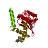
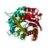
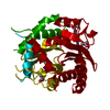
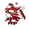
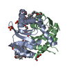
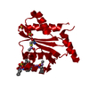
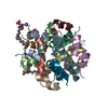
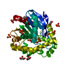
| Title | HIV INTEGRASE CORE DOMAIN COMPLEXED WITH A DERIVATIVE OF TETRAPHENYL ARSONIUM. | ||||||
 Components Components | INTEGRASE | ||||||
 Keywords Keywords | TRANSFERASE / DNA INTEGRATION | ||||||
| Function / homology |  Function and homology information Function and homology informationHIV-1 retropepsin / symbiont-mediated activation of host apoptosis / retroviral ribonuclease H / exoribonuclease H / exoribonuclease H activity / DNA integration / viral genome integration into host DNA / establishment of integrated proviral latency / RNA-directed DNA polymerase / RNA stem-loop binding ...HIV-1 retropepsin / symbiont-mediated activation of host apoptosis / retroviral ribonuclease H / exoribonuclease H / exoribonuclease H activity / DNA integration / viral genome integration into host DNA / establishment of integrated proviral latency / RNA-directed DNA polymerase / RNA stem-loop binding / viral penetration into host nucleus / host multivesicular body / RNA-directed DNA polymerase activity / RNA-DNA hybrid ribonuclease activity / Transferases; Transferring phosphorus-containing groups; Nucleotidyltransferases / host cell / viral nucleocapsid / endonuclease activity / DNA recombination / DNA-directed DNA polymerase / aspartic-type endopeptidase activity / Hydrolases; Acting on ester bonds / host cell cytoplasm / DNA-directed DNA polymerase activity / symbiont-mediated suppression of host gene expression / viral translational frameshifting / symbiont entry into host cell / lipid binding / host cell nucleus / host cell plasma membrane / virion membrane / structural molecule activity / proteolysis / DNA binding / zinc ion binding Similarity search - Function | ||||||
| Biological species |   Human immunodeficiency virus 1 Human immunodeficiency virus 1 | ||||||
| Method |  X-RAY DIFFRACTION / MOLECULAR REPLACEMENT / Resolution: 2.3 Å X-RAY DIFFRACTION / MOLECULAR REPLACEMENT / Resolution: 2.3 Å | ||||||
 Authors Authors | Molteni, V. / Greenwald, J. / Rhodes, D. / Hwang, Y. / Kwiatkowski, W. / Bushman, F.D. / Siegel, J.S. / Choe, S. | ||||||
 Citation Citation | Journal: Acta Crystallogr.,Sect.D / Year: 2001 Title: Identification of a small-molecule binding site at the dimer interface of the HIV integrase catalytic domain. Authors: Molteni, V. / Greenwald, J. / Rhodes, D. / Hwang, Y. / Kwiatkowski, W. / Bushman, F.D. / Siegel, J.S. / Choe, S. | ||||||
| History |
| ||||||
| Remark 999 | Sequence The protein was expressed with N-term 6x histidine tag which when removed with thrombin ...Sequence The protein was expressed with N-term 6x histidine tag which when removed with thrombin leaves GSH at the N-terminus. |
- Structure visualization
| Structure viewer | Molecule: MolmilJmol/JSmol |
|---|
- Downloads & links
-Download
| PDBx/mmCIF format | 1hyz.cif.gz | 46.6 KB | Display | PDBx/mmCIF format |
|---|---|---|---|---|
| PDB format | pdb1hyz.ent.gz | 31.4 KB | Display | PDB format |
| PDBx/mmJSON format | 1hyz.json.gz | Tree view | PDBx/mmJSON format | |
| Others |  Other downloads Other downloads |
-Validation report
| Arichive directory | https://data.pdbj.org/pub/pdb/validation_reports/hy/1hyzftp://data.pdbj.org/pub/pdb/validation_reports/hy/1hyz | HTTPS FTP |
|---|
-Related structure data

-Links
 PDBj
PDBj
- Assembly
| Deposited unit | 
| ||||||||
|---|---|---|---|---|---|---|---|---|---|
| 1 | 
| ||||||||
| Unit cell |
| ||||||||
| Details | Crystallographic 2-fold creates biologically relevant dimer |
-Components
| #1: Protein | Mass: 18434.723 Da / Num. of mol.: 1 / Fragment: CATALYTIC CORE DOMAIN (RESIDUES 50-212) / Mutation: F185K Source method: isolated from a genetically manipulated source Source: (gene. exp.) Human immunodeficiency virus 1 / Genus: Lentivirus / Production host:  References: UniProt: Q76353, UniProt: P12497*PLUS, RNA-directed DNA polymerase |
|---|---|
| #2: Chemical | ChemComp-SO4 /   Mass: 96.063 Da / Num. of mol.: 1 / Source method: obtained synthetically / Formula: SO4 Mass: 96.063 Da / Num. of mol.: 1 / Source method: obtained synthetically / Formula: SO4 |
| #3: Chemical | ChemComp-CL /   Mass: 35.453 Da / Num. of mol.: 1 / Source method: obtained synthetically / Formula: Cl Mass: 35.453 Da / Num. of mol.: 1 / Source method: obtained synthetically / Formula: Cl |
| #4: Chemical | ChemComp-TTO / (  Mass: 415.336 Da / Num. of mol.: 1 / Source method: obtained synthetically / Formula: C24H20AsO2 Mass: 415.336 Da / Num. of mol.: 1 / Source method: obtained synthetically / Formula: C24H20AsO2 |
| #5: Water | ChemComp-HOH /  Mass: 18.015 Da / Num. of mol.: 81 / Source method: isolated from a natural source / Formula: H2O Mass: 18.015 Da / Num. of mol.: 81 / Source method: isolated from a natural source / Formula: H2O |
| Has protein modification | Y |
-Experimental details
-Experiment
| Experiment | Method: X-RAY DIFFRACTION / Number of used crystals: 1 |
|---|
- Sample preparation
| Crystal | Density Matthews: 2.66 Å3/Da / Density % sol: 53.76 % | ||||||||||||||||||||||||||||||||||||||||||||||||
|---|---|---|---|---|---|---|---|---|---|---|---|---|---|---|---|---|---|---|---|---|---|---|---|---|---|---|---|---|---|---|---|---|---|---|---|---|---|---|---|---|---|---|---|---|---|---|---|---|---|
| Crystal grow | Temperature: 277 K / Method: vapor diffusion, hanging drop / pH: 6.5 Details: PEG8000, NH4SO4, Cacodylate, pH 6.5, VAPOR DIFFUSION, HANGING DROP, temperature 277K | ||||||||||||||||||||||||||||||||||||||||||||||||
| Crystal grow | *PLUS | ||||||||||||||||||||||||||||||||||||||||||||||||
| Components of the solutions | *PLUS
|
-Data collection
| Diffraction | Mean temperature: 100 K |
|---|---|
| Diffraction source | Source: ROTATING ANODE / Type: OTHER / Wavelength: 1.54 Å |
| Detector | Type: MACSCIENCE / Detector: IMAGE PLATE / Date: May 1, 1997 / Details: focusing mirror |
| Radiation | Protocol: SINGLE WAVELENGTH / Monochromatic (M) / Laue (L): M / Scattering type: x-ray |
| Radiation wavelength | Wavelength: 1.54 Å / Relative weight: 1 |
| Reflection | Resolution: 2.3→25 Å / Num. all: 15753 / Num. obs: 15753 / % possible obs: 92.7 % / Observed criterion σ(F): 0 / Observed criterion σ(I): 0 / Redundancy: 11 % / Biso Wilson estimate: 48 Å2 / Rmerge(I) obs: 0.043 / Net I/σ(I): 24 |
| Reflection shell | Resolution: 2.3→2.38 Å / Rmerge(I) obs: 0.288 / % possible all: 92.6 |
| Reflection shell | *PLUS % possible obs: 92.6 % |
- Processing
| Software |
| ||||||||||||||||||||||||||||||
|---|---|---|---|---|---|---|---|---|---|---|---|---|---|---|---|---|---|---|---|---|---|---|---|---|---|---|---|---|---|---|---|
| Refinement | Method to determine structure: MOLECULAR REPLACEMENT / Resolution: 2.3→24 Å / σ(F): 0 / σ(I): 0 / Stereochemistry target values: default for refmac
| ||||||||||||||||||||||||||||||
| Refinement step | Cycle: LAST / Resolution: 2.3→24 Å
| ||||||||||||||||||||||||||||||
| Software | *PLUS Name: REFMAC / Classification: refinement | ||||||||||||||||||||||||||||||
| Refine LS restraints | *PLUS
|
