 Movie
Movie Controller

[English] 日本語
 Yorodumi
- PDB-8p7f: The impact of molecular variants, crystallization conditions and ... -
+ Open data

- Basic information
| Entry | Database: PDB / ID: 8p7f | |||||||||
|---|---|---|---|---|---|---|---|---|---|---|
| Title | The impact of molecular variants, crystallization conditions and space group on structure-ligand complexes: A case study on Bacterial Phosphotriesterase Variants and complexes | |||||||||
 Components Components | Parathion hydrolase | |||||||||
 Keywords Keywords | HYDROLASE | |||||||||
| Function / homology |  Function and homology information Function and homology informationaryldialkylphosphatase / aryldialkylphosphatase activity / zinc ion binding / plasma membrane Similarity search - Function | |||||||||
| Biological species |  Brevundimonas diminuta (bacteria) Brevundimonas diminuta (bacteria) | |||||||||
| Method |  X-RAY DIFFRACTION / SYNCHROTRON / MOLECULAR REPLACEMENT / Resolution: 2 Å X-RAY DIFFRACTION / SYNCHROTRON / MOLECULAR REPLACEMENT / Resolution: 2 Å | |||||||||
 Authors Authors | Dym, O. / Aggarwal, N. / Ashani, Y. / Albeck, S. / Unger, T. / Hamer Rogotner, S. / Silman, I. / Sussman, J.L. | |||||||||
| Funding support |  Israel, 1items Israel, 1items
| |||||||||
 Citation Citation | Journal: Acta Crystallogr D Struct Biol / Year: 2023 Title: The impact of molecular variants, crystallization conditions and the space group on ligand-protein complexes: a case study on bacterial phosphotriesterase. Authors: Dym, O. / Aggarwal, N. / Ashani, Y. / Leader, H. / Albeck, S. / Unger, T. / Hamer-Rogotner, S. / Silman, I. / Tawfik, D.S. / Sussman, J.L. | |||||||||
| History |
|
- Structure visualization
| Structure viewer | Molecule: MolmilJmol/JSmol |
|---|
- Downloads & links
-Download
| PDBx/mmCIF format | 8p7f.cif.gz | 73.2 KB | Display | PDBx/mmCIF format |
|---|---|---|---|---|
| PDB format | pdb8p7f.ent.gz | 50.1 KB | Display | PDB format |
| PDBx/mmJSON format | 8p7f.json.gz | Tree view | PDBx/mmJSON format | |
| Others |  Other downloads Other downloads |
-Validation report
| Arichive directory | https://data.pdbj.org/pub/pdb/validation_reports/p7/8p7fftp://data.pdbj.org/pub/pdb/validation_reports/p7/8p7f | HTTPS FTP |
|---|
-Related structure data
| Related structure data |  8p7hC  8p7iC  8p7kC  8p7mC  8p7nC  8p7qC  8p7rC 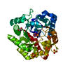 8p7sC  8p7tC  8p7uC  8p7vC C: citing same article ( |
|---|---|
| Similar structure data |
-Links
 PDBj
PDBj


- Assembly
| Deposited unit | 
| ||||||||
|---|---|---|---|---|---|---|---|---|---|
| 1 |
| ||||||||
| Unit cell |
|
-Components
| #1: Protein | Mass: 35864.684 Da / Num. of mol.: 1 Source method: isolated from a genetically manipulated source Source: (gene. exp.) Brevundimonas diminuta (bacteria) / Gene: opd / Production host: |
|---|---|
| #2: Chemical | ChemComp-ZN /   Mass: 65.409 Da / Num. of mol.: 1 / Source method: obtained synthetically / Formula: Zn / Feature type: SUBJECT OF INVESTIGATION Mass: 65.409 Da / Num. of mol.: 1 / Source method: obtained synthetically / Formula: Zn / Feature type: SUBJECT OF INVESTIGATION |
| #3: Water | ChemComp-HOH /  Mass: 18.015 Da / Num. of mol.: 84 / Source method: isolated from a natural source / Formula: H2O Mass: 18.015 Da / Num. of mol.: 84 / Source method: isolated from a natural source / Formula: H2O |
| Has ligand of interest | Y |
-Experimental details
-Experiment
| Experiment | Method: X-RAY DIFFRACTION / Number of used crystals: 1 |
|---|
- Sample preparation
| Crystal | Density Matthews: 2.32 Å3/Da / Density % sol: 46.99 % |
|---|---|
| Crystal grow | Temperature: 292 K / Method: vapor diffusion, hanging drop / pH: 7.5 / Details: 12% PEG 6,000 5% MPD 0.1M HEPES pH=7.5 |
-Data collection
| Diffraction | Mean temperature: 100 K / Serial crystal experiment: N |
|---|---|
| Diffraction source | Source: SYNCHROTRON / Site: ESRF  / Beamline: ID14-4 / Wavelength: 0.9763 Å / Beamline: ID14-4 / Wavelength: 0.9763 Å |
| Detector | Type: MARMOSAIC 300 mm CCD / Detector: CCD / Date: Nov 25, 2013 |
| Radiation | Protocol: SINGLE WAVELENGTH / Monochromatic (M) / Laue (L): M / Scattering type: x-ray |
| Radiation wavelength | Wavelength: 0.9763 Å / Relative weight: 1 |
| Reflection | Resolution: 2→40.261 Å / Num. obs: 23037 / % possible obs: 98.91 % / Redundancy: 5.3 % / CC1/2: 0.75 / Net I/σ(I): 12.5 |
| Reflection shell | Resolution: 2→2.071 Å / Num. unique obs: 2273 / CC1/2: 0.638 |
- Processing
| Software |
| |||||||||||||||||||||||||||||||||||||||||||||||||||||||||||||||
|---|---|---|---|---|---|---|---|---|---|---|---|---|---|---|---|---|---|---|---|---|---|---|---|---|---|---|---|---|---|---|---|---|---|---|---|---|---|---|---|---|---|---|---|---|---|---|---|---|---|---|---|---|---|---|---|---|---|---|---|---|---|---|---|---|
| Refinement | Method to determine structure: MOLECULAR REPLACEMENT / Resolution: 2→40.261 Å / SU ML: 0.19 / Cross valid method: FREE R-VALUE / σ(F): 1.36 / Phase error: 25.02 / Stereochemistry target values: ML
| |||||||||||||||||||||||||||||||||||||||||||||||||||||||||||||||
| Solvent computation | Shrinkage radii: 0.9 Å / VDW probe radii: 1.11 Å / Solvent model: FLAT BULK SOLVENT MODEL | |||||||||||||||||||||||||||||||||||||||||||||||||||||||||||||||
| Refinement step | Cycle: LAST / Resolution: 2→40.261 Å
| |||||||||||||||||||||||||||||||||||||||||||||||||||||||||||||||
| Refine LS restraints |
| |||||||||||||||||||||||||||||||||||||||||||||||||||||||||||||||
| LS refinement shell |
|
