 Movie
Movie Controller

[English] 日本語
 Yorodumi
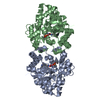
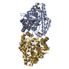
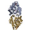
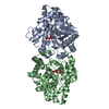
- PDB-1eyw: THREE-DIMENSIONAL STRUCTURE OF THE ZINC-CONTAINING PHOSPHOTRIESTE... -
+ Open data

- Basic information
| Entry | Database: PDB / ID: 1eyw | ||||||
|---|---|---|---|---|---|---|---|
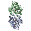
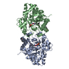
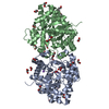
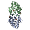
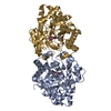
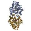
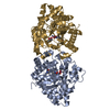
| Title | THREE-DIMENSIONAL STRUCTURE OF THE ZINC-CONTAINING PHOSPHOTRIESTERASE WITH BOUND SUBSTRATE ANALOG TRIETHYLPHOSPHATE | ||||||
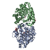
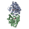
 Components Components | PHOSPHOTRIESTERASE | ||||||
 Keywords Keywords | HYDROLASE / ORGANOPHOSPHATE / ZINC | ||||||
| Function / homology |  Function and homology information Function and homology informationaryldialkylphosphatase / aryldialkylphosphatase activity / zinc ion binding / plasma membrane Similarity search - Function | ||||||
| Biological species |  Brevundimonas diminuta (bacteria) Brevundimonas diminuta (bacteria) | ||||||
| Method |  X-RAY DIFFRACTION / Resolution: 1.9 Å X-RAY DIFFRACTION / Resolution: 1.9 Å | ||||||
 Authors Authors | Holden, H.M. / Benning, M.M. / Raushel, F.M. / Hong, S.-B. | ||||||
 Citation Citation | Journal: J.Biol.Chem. / Year: 2000 Title: The binding of substrate analogs to phosphotriesterase. Authors: Benning, M.M. / Hong, S.B. / Raushel, F.M. / Holden, H.M. | ||||||
| History |
|
- Structure visualization
| Structure viewer | Molecule: MolmilJmol/JSmol |
|---|
- Downloads & links
-Download
| PDBx/mmCIF format | 1eyw.cif.gz | 83 KB | Display | PDBx/mmCIF format |
|---|---|---|---|---|
| PDB format | pdb1eyw.ent.gz | 60 KB | Display | PDB format |
| PDBx/mmJSON format | 1eyw.json.gz | Tree view | PDBx/mmJSON format | |
| Others |  Other downloads Other downloads |
-Validation report
| Arichive directory | https://data.pdbj.org/pub/pdb/validation_reports/ey/1eywftp://data.pdbj.org/pub/pdb/validation_reports/ey/1eyw | HTTPS FTP |
|---|
-Related structure data


-Links
 PDBj
PDBj


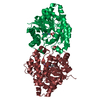
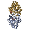
- Assembly
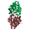
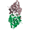
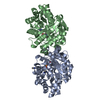
| Deposited unit | 
| ||||||||
|---|---|---|---|---|---|---|---|---|---|
| 1 | 
| ||||||||
| Unit cell |
|
-Components
| #1: Protein | Mass: 35959.961 Da / Num. of mol.: 1 Source method: isolated from a genetically manipulated source Source: (gene. exp.) Brevundimonas diminuta (bacteria) / Plasmid: PKK01 / Production host: | ||||||||
|---|---|---|---|---|---|---|---|---|---|
| #2: Chemical |   Mass: 65.409 Da / Num. of mol.: 2 / Source method: obtained synthetically / Formula: Zn Mass: 65.409 Da / Num. of mol.: 2 / Source method: obtained synthetically / Formula: Zn#3: Chemical | ChemComp-TEN / |   Mass: 182.155 Da / Num. of mol.: 1 / Source method: obtained synthetically / Formula: C6H15O4P Mass: 182.155 Da / Num. of mol.: 1 / Source method: obtained synthetically / Formula: C6H15O4P#4: Chemical | ChemComp-PEL / | 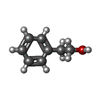  Mass: 122.164 Da / Num. of mol.: 1 / Source method: obtained synthetically / Formula: C8H10O Mass: 122.164 Da / Num. of mol.: 1 / Source method: obtained synthetically / Formula: C8H10O#5: Water | ChemComp-HOH / |  Mass: 18.015 Da / Num. of mol.: 221 / Source method: isolated from a natural source / Formula: H2O Mass: 18.015 Da / Num. of mol.: 221 / Source method: isolated from a natural source / Formula: H2OHas protein modification | Y | |
-Experimental details
-Experiment
| Experiment | Method: X-RAY DIFFRACTION / Number of used crystals: 1 |
|---|
- Sample preparation
| Crystal | Density Matthews: 2.88 Å3/Da / Density % sol: 57.33 % | ||||||||||||||||||||||||||||||||||||||||||
|---|---|---|---|---|---|---|---|---|---|---|---|---|---|---|---|---|---|---|---|---|---|---|---|---|---|---|---|---|---|---|---|---|---|---|---|---|---|---|---|---|---|---|---|
| Crystal grow | Temperature: 277 K / Method: batch / pH: 9 Details: PEG 8000, Sodium Chloride, CHES, pH 9.0, batch, temperature 277K | ||||||||||||||||||||||||||||||||||||||||||
| Crystal | *PLUS Density % sol: 56 % | ||||||||||||||||||||||||||||||||||||||||||
| Crystal grow | *PLUS Temperature: 4 ℃ / Method: batch method / Details: macroseeding | ||||||||||||||||||||||||||||||||||||||||||
| Components of the solutions | *PLUS
|
-Data collection
| Diffraction | Mean temperature: 277 K |
|---|---|
| Diffraction source | Source: ROTATING ANODE / Type: RIGAKU RU200 / Wavelength: 1.5418 |
| Detector | Type: SIEMENS HI-STAR / Detector: AREA DETECTOR / Date: Oct 15, 1997 |
| Radiation | Protocol: SINGLE WAVELENGTH / Monochromatic (M) / Laue (L): M / Scattering type: x-ray |
| Radiation wavelength | Wavelength: 1.5418 Å / Relative weight: 1 |
| Reflection | Resolution: 1.9→20 Å / Num. obs: 29135 / % possible obs: 92 % / Observed criterion σ(I): 0 / Redundancy: 4.3 % / Rmerge(I) obs: 0.046 / Net I/σ(I): 18 |
| Reflection shell | Resolution: 1.9→1.99 Å / Redundancy: 2.2 % / Rmerge(I) obs: 0.162 / % possible all: 74 |
| Reflection shell | *PLUS % possible obs: 74 % |
- Processing
| Software |
| ||||||||||||||||||||||||||||||
|---|---|---|---|---|---|---|---|---|---|---|---|---|---|---|---|---|---|---|---|---|---|---|---|---|---|---|---|---|---|---|---|
| Refinement | Resolution: 1.9→20 Å / σ(F): 0 / Stereochemistry target values: TNT STANDARD GEOMETRY
| ||||||||||||||||||||||||||||||
| Refinement step | Cycle: LAST / Resolution: 1.9→20 Å
| ||||||||||||||||||||||||||||||
| Refine LS restraints |
| ||||||||||||||||||||||||||||||
| Software | *PLUS Name: TNT / Version: 5E / Classification: refinement | ||||||||||||||||||||||||||||||
| Refinement | *PLUS Rfactor Rfree: 0.211 | ||||||||||||||||||||||||||||||
| Solvent computation | *PLUS | ||||||||||||||||||||||||||||||
| Displacement parameters | *PLUS | ||||||||||||||||||||||||||||||
| Refine LS restraints | *PLUS
|
