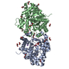
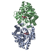
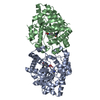
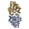
 Movie
Movie Controller


+ Open data

- Basic information
| Entry | Database: PDB / ID: 4xd3 | ||||||
|---|---|---|---|---|---|---|---|
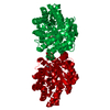
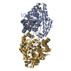
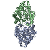
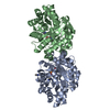
| Title | Phosphotriesterase variant E3 | ||||||
 Components Components | Phosphotriesterase variant PTE-E1 | ||||||
 Keywords Keywords | HYDROLASE / dynamics / evolution / phosphotriesterase | ||||||
| Function / homology |  Function and homology information Function and homology informationcatabolic process / hydrolase activity, acting on ester bonds / zinc ion binding Similarity search - Function | ||||||
| Biological species |  Brevundimonas diminuta (bacteria) Brevundimonas diminuta (bacteria) | ||||||
| Method |  X-RAY DIFFRACTION / SYNCHROTRON / MOLECULAR REPLACEMENT / Resolution: 1.57 Å X-RAY DIFFRACTION / SYNCHROTRON / MOLECULAR REPLACEMENT / Resolution: 1.57 Å | ||||||
 Authors Authors | Jackson, C.J. / Campbell, E. / Kaltenbach, M. / Tokuriki, N. | ||||||
 Citation Citation | Journal: Nat.Chem.Biol. / Year: 2016 Title: The role of protein dynamics in the evolution of new enzyme function. Authors: Campbell, E. / Kaltenbach, M. / Correy, G.J. / Carr, P.D. / Porebski, B.T. / Livingstone, E.K. / Afriat-Jurnou, L. / Buckle, A.M. / Weik, M. / Hollfelder, F. / Tokuriki, N. / Jackson, C.J. | ||||||
| History |
|
- Structure visualization
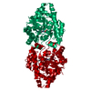
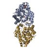
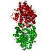
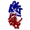
| Structure viewer | Molecule: MolmilJmol/JSmol |
|---|
- Downloads & links
-Download
| PDBx/mmCIF format | 4xd3.cif.gz | 159.2 KB | Display | PDBx/mmCIF format |
|---|---|---|---|---|
| PDB format | pdb4xd3.ent.gz | 122 KB | Display | PDB format |
| PDBx/mmJSON format | 4xd3.json.gz | Tree view | PDBx/mmJSON format | |
| Others |  Other downloads Other downloads |
-Validation report
| Arichive directory | https://data.pdbj.org/pub/pdb/validation_reports/xd/4xd3ftp://data.pdbj.org/pub/pdb/validation_reports/xd/4xd3 | HTTPS FTP |
|---|
-Related structure data
| Related structure data |  4pbeC  4pbfC  4pcnC  4pcpC  4xafC  4xagC  4xayC  4xazC  4xd4C  4xd5C  4xd6C  4gy0S C: citing same article ( S: Starting model for refinement |
|---|---|
| Similar structure data |


-Links
 PDBj
PDBj


- Assembly
| Deposited unit | 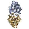
| ||||||||
|---|---|---|---|---|---|---|---|---|---|
| 1 |
| ||||||||
| Unit cell |
| ||||||||
| Components on special symmetry positions |
|
-Components
| #1: Protein | Mass: 36141.258 Da / Num. of mol.: 2 Source method: isolated from a genetically manipulated source Source: (gene. exp.) Brevundimonas diminuta (bacteria) / Production host: #2: Chemical | ChemComp-ZN /   Mass: 65.409 Da / Num. of mol.: 4 / Source method: obtained synthetically / Formula: Zn Mass: 65.409 Da / Num. of mol.: 4 / Source method: obtained synthetically / Formula: Zn#3: Chemical |   Mass: 136.989 Da / Num. of mol.: 2 / Source method: obtained synthetically / Formula: C2H6AsO2 Mass: 136.989 Da / Num. of mol.: 2 / Source method: obtained synthetically / Formula: C2H6AsO2#4: Chemical | ChemComp-MPD / (   Mass: 118.174 Da / Num. of mol.: 4 / Source method: obtained synthetically / Formula: C6H14O2 / Comment: precipitant*YM Mass: 118.174 Da / Num. of mol.: 4 / Source method: obtained synthetically / Formula: C6H14O2 / Comment: precipitant*YM#5: Water | ChemComp-HOH / |  Mass: 18.015 Da / Num. of mol.: 672 / Source method: isolated from a natural source / Formula: H2O Mass: 18.015 Da / Num. of mol.: 672 / Source method: isolated from a natural source / Formula: H2O |
|---|
-Experimental details
-Experiment
| Experiment | Method: X-RAY DIFFRACTION / Number of used crystals: 1 |
|---|
- Sample preparation
| Crystal | Density Matthews: 2.26 Å3/Da / Density % sol: 45.62 % |
|---|---|
| Crystal grow | Temperature: 277.15 K / Method: vapor diffusion, hanging drop / Details: 100 mM NaCacodylate 30% 2-methane-2,4-pentanediol / PH range: 6-7 |
-Data collection
| Diffraction | Mean temperature: 100 K |
|---|---|
| Diffraction source | Source: SYNCHROTRON / Site: Australian Synchrotron  / Beamline: MX1 / Wavelength: 0.9537 Å / Beamline: MX1 / Wavelength: 0.9537 Å |
| Detector | Type: ADSC QUANTUM 210r / Detector: CCD / Date: Apr 18, 2014 |
| Radiation | Protocol: SINGLE WAVELENGTH / Monochromatic (M) / Laue (L): M / Scattering type: x-ray |
| Radiation wavelength | Wavelength: 0.9537 Å / Relative weight: 1 |
| Reflection | Resolution: 1.57→39.41 Å / Num. obs: 91982 / % possible obs: 99.91 % / Redundancy: 7.3 % / Rmerge(I) obs: 0.0828 / Net I/σ(I): 16.45 |
| Reflection shell | Resolution: 1.57→1.626 Å / Redundancy: 7.3 % / Rmerge(I) obs: 0.9643 / Mean I/σ(I) obs: 2.31 / % possible all: 99.94 |
- Processing
| Software |
| |||||||||||||||||||||||||||||||||||||||||||||||||||||||||||||||||||||||||||||||||||||||||||||||||||||||||||||||||||||||||||||||||||||||||||||||||||||||||||||||||||||||||||||||||||||||||||||||||||||||||||||||||||||||||
|---|---|---|---|---|---|---|---|---|---|---|---|---|---|---|---|---|---|---|---|---|---|---|---|---|---|---|---|---|---|---|---|---|---|---|---|---|---|---|---|---|---|---|---|---|---|---|---|---|---|---|---|---|---|---|---|---|---|---|---|---|---|---|---|---|---|---|---|---|---|---|---|---|---|---|---|---|---|---|---|---|---|---|---|---|---|---|---|---|---|---|---|---|---|---|---|---|---|---|---|---|---|---|---|---|---|---|---|---|---|---|---|---|---|---|---|---|---|---|---|---|---|---|---|---|---|---|---|---|---|---|---|---|---|---|---|---|---|---|---|---|---|---|---|---|---|---|---|---|---|---|---|---|---|---|---|---|---|---|---|---|---|---|---|---|---|---|---|---|---|---|---|---|---|---|---|---|---|---|---|---|---|---|---|---|---|---|---|---|---|---|---|---|---|---|---|---|---|---|---|---|---|---|---|---|---|---|---|---|---|---|---|---|---|---|---|---|---|---|
| Refinement | Method to determine structure: MOLECULAR REPLACEMENT Starting model: 4GY0 Resolution: 1.57→39.408 Å / SU ML: 0.17 / Cross valid method: FREE R-VALUE / σ(F): 1.34 / Phase error: 21.15 / Stereochemistry target values: ML
| |||||||||||||||||||||||||||||||||||||||||||||||||||||||||||||||||||||||||||||||||||||||||||||||||||||||||||||||||||||||||||||||||||||||||||||||||||||||||||||||||||||||||||||||||||||||||||||||||||||||||||||||||||||||||
| Solvent computation | Shrinkage radii: 0.9 Å / VDW probe radii: 1.11 Å / Solvent model: FLAT BULK SOLVENT MODEL | |||||||||||||||||||||||||||||||||||||||||||||||||||||||||||||||||||||||||||||||||||||||||||||||||||||||||||||||||||||||||||||||||||||||||||||||||||||||||||||||||||||||||||||||||||||||||||||||||||||||||||||||||||||||||
| Refinement step | Cycle: LAST / Resolution: 1.57→39.408 Å
| |||||||||||||||||||||||||||||||||||||||||||||||||||||||||||||||||||||||||||||||||||||||||||||||||||||||||||||||||||||||||||||||||||||||||||||||||||||||||||||||||||||||||||||||||||||||||||||||||||||||||||||||||||||||||
| Refine LS restraints |
| |||||||||||||||||||||||||||||||||||||||||||||||||||||||||||||||||||||||||||||||||||||||||||||||||||||||||||||||||||||||||||||||||||||||||||||||||||||||||||||||||||||||||||||||||||||||||||||||||||||||||||||||||||||||||
| LS refinement shell |
|
