 Movie
Movie Controller

[English] 日本語
 Yorodumi
- PDB-3urq: Crystal Structure of PTE mutant H254G/H257W/L303T/M317L/I106C/F13... -
+ Open data

- Basic information
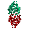
| Entry | Database: PDB / ID: 3urq | ||||||
|---|---|---|---|---|---|---|---|
| Title | Crystal Structure of PTE mutant H254G/H257W/L303T/M317L/I106C/F132I/L271I/K185R/I274N/A80V/R67H with cyclohexyl methylphosphonate inhibitor | ||||||
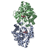
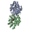
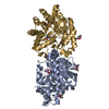
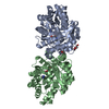
 Components Components | Parathion hydrolase | ||||||
 Keywords Keywords | HYDROLASE/HYDROLASE INHIBITOR / metalloenzyme / TIM barrel / nerve agents / phosphotriesterase / HYDROLASE / HYDROLASE-HYDROLASE INHIBITOR complex | ||||||
| Function / homology |  Function and homology information Function and homology informationaryldialkylphosphatase / aryldialkylphosphatase activity / zinc ion binding / plasma membrane Similarity search - Function | ||||||
| Biological species |  Brevundimonas diminuta (bacteria) Brevundimonas diminuta (bacteria) | ||||||
| Method |  X-RAY DIFFRACTION / SYNCHROTRON / MOLECULAR REPLACEMENT / Resolution: 2.1 Å X-RAY DIFFRACTION / SYNCHROTRON / MOLECULAR REPLACEMENT / Resolution: 2.1 Å | ||||||
 Authors Authors | Tsai, P. / Fox, N.G. / Li, Y. / Barondeau, D.P. / Raushel, F.M. | ||||||
 Citation Citation | Journal: Biochemistry / Year: 2012 Title: Enzymes for the homeland defense: optimizing phosphotriesterase for the hydrolysis of organophosphate nerve agents. Authors: Tsai, P.C. / Fox, N. / Bigley, A.N. / Harvey, S.P. / Barondeau, D.P. / Raushel, F.M. | ||||||
| History |
|
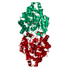
- Structure visualization
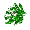
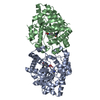
| Structure viewer | Molecule: MolmilJmol/JSmol |
|---|
- Downloads & links
-Download
| PDBx/mmCIF format | 3urq.cif.gz | 144.9 KB | Display | PDBx/mmCIF format |
|---|---|---|---|---|
| PDB format | pdb3urq.ent.gz | 111.6 KB | Display | PDB format |
| PDBx/mmJSON format | 3urq.json.gz | Tree view | PDBx/mmJSON format | |
| Others |  Other downloads Other downloads |
-Validation report
| Arichive directory | https://data.pdbj.org/pub/pdb/validation_reports/ur/3urqftp://data.pdbj.org/pub/pdb/validation_reports/ur/3urq | HTTPS FTP |
|---|
-Related structure data
| Related structure data |  3upmC  3ur2C  3ur5C  3uraC 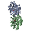 3urbSC 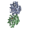 3urnC C: citing same article ( S: Starting model for refinement |
|---|---|
| Similar structure data |


-Links
 PDBj
PDBj


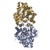
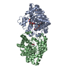
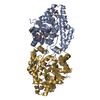
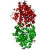
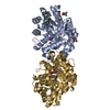
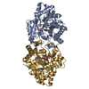
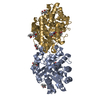
- Assembly
| Deposited unit | 
| |||||||||
|---|---|---|---|---|---|---|---|---|---|---|
| 1 |
| |||||||||
| Unit cell |
| |||||||||
| Components on special symmetry positions |
|
-Components
| #1: Protein | Mass: 35462.254 Da / Num. of mol.: 2 / Fragment: UNP residues 35-361 Mutation: H254G/H257W/L303T/M317L/I106C/F132I/L271I/K185R/I274N/A80V/R67H Source method: isolated from a genetically manipulated source Source: (gene. exp.) Brevundimonas diminuta (bacteria) / Gene: opd / Production host: #2: Chemical | ChemComp-CO /   Mass: 58.933 Da / Num. of mol.: 4 / Source method: obtained synthetically / Formula: Co Mass: 58.933 Da / Num. of mol.: 4 / Source method: obtained synthetically / Formula: Co#3: Chemical |   Mass: 178.166 Da / Num. of mol.: 2 / Source method: obtained synthetically / Formula: C7H15O3P Mass: 178.166 Da / Num. of mol.: 2 / Source method: obtained synthetically / Formula: C7H15O3P#4: Chemical | ChemComp-IMD /   Mass: 69.085 Da / Num. of mol.: 4 / Source method: obtained synthetically / Formula: C3H5N2 Mass: 69.085 Da / Num. of mol.: 4 / Source method: obtained synthetically / Formula: C3H5N2#5: Water | ChemComp-HOH / |  Mass: 18.015 Da / Num. of mol.: 316 / Source method: isolated from a natural source / Formula: H2O Mass: 18.015 Da / Num. of mol.: 316 / Source method: isolated from a natural source / Formula: H2O |
|---|
-Experimental details
-Experiment
| Experiment | Method: X-RAY DIFFRACTION / Number of used crystals: 1 |
|---|
- Sample preparation
| Crystal | Density Matthews: 2.26 Å3/Da / Density % sol: 45.53 % |
|---|---|
| Crystal grow | Temperature: 295 K / Method: vapor diffusion, hanging drop / pH: 7 Details: 2 uL 7.8 mg/mL protein + 2 uL seeding solution (24% PEG5000 MME, 1.0 mM cobalt chloride, 100 mM cyclohexyl methylphosphonate, 0.1 M imidazole, pH 7.0) over 500 uL precipitating agent (26% ...Details: 2 uL 7.8 mg/mL protein + 2 uL seeding solution (24% PEG5000 MME, 1.0 mM cobalt chloride, 100 mM cyclohexyl methylphosphonate, 0.1 M imidazole, pH 7.0) over 500 uL precipitating agent (26% PEG5000 MME), VAPOR DIFFUSION, HANGING DROP, temperature 295K |
-Data collection
| Diffraction | Mean temperature: 100 K |
|---|---|
| Diffraction source | Source: SYNCHROTRON / Site: SSRL  / Beamline: BL7-1 / Wavelength: 0.9794 Å / Beamline: BL7-1 / Wavelength: 0.9794 Å |
| Detector | Type: ADSC QUANTUM 315r / Detector: CCD / Date: Jan 29, 2010 |
| Radiation | Monochromator: Si(111), side scattering I-beam bent single crystal, asymmetric cut 4.9650 deg Protocol: SINGLE WAVELENGTH / Monochromatic (M) / Laue (L): M / Scattering type: x-ray |
| Radiation wavelength | Wavelength: 0.9794 Å / Relative weight: 1 |
| Reflection | Resolution: 2.1→50 Å / Num. all: 38049 / Num. obs: 38029 / % possible obs: 99.9 % / Observed criterion σ(F): 0 / Observed criterion σ(I): 0 / Redundancy: 8 % / Biso Wilson estimate: 31 Å2 / Rsym value: 0.0066 / Net I/σ(I): 23.3 |
| Reflection shell | Resolution: 2.1→2.18 Å / Redundancy: 8.1 % / Mean I/σ(I) obs: 6 / Num. unique all: 3726 / Rsym value: 0.395 / % possible all: 100 |
- Processing
| Software |
| ||||||||||||||||||||||||||||
|---|---|---|---|---|---|---|---|---|---|---|---|---|---|---|---|---|---|---|---|---|---|---|---|---|---|---|---|---|---|
| Refinement | Method to determine structure: MOLECULAR REPLACEMENT Starting model: PDB ENTRY 3URB Resolution: 2.1→50 Å / Occupancy max: 1 / Occupancy min: 1 / Cross valid method: THROUGHOUT / σ(F): 0 / Stereochemistry target values: Engh & Huber
| ||||||||||||||||||||||||||||
| Solvent computation | Bsol: 15.1491 Å2 | ||||||||||||||||||||||||||||
| Displacement parameters | Biso max: 97.11 Å2 / Biso mean: 38.2081 Å2 / Biso min: 16.49 Å2
| ||||||||||||||||||||||||||||
| Refinement step | Cycle: LAST / Resolution: 2.1→50 Å
| ||||||||||||||||||||||||||||
| Refine LS restraints |
| ||||||||||||||||||||||||||||
| LS refinement shell | Resolution: 2.1→2.18 Å
| ||||||||||||||||||||||||||||
| Xplor file |
|
