 Movie
Movie Controller

[English] 日本語
 Yorodumi
- PDB-1p6c: crystal structure of phosphotriesterase triple mutant H254G/H257W... -
+ Open data

- Basic information
| Entry | Database: PDB / ID: 1p6c | ||||||
|---|---|---|---|---|---|---|---|
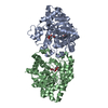
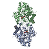
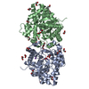
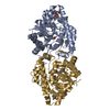
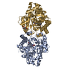
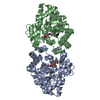
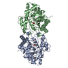
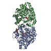
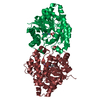
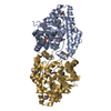
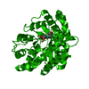
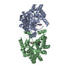
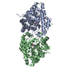
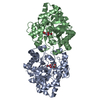
| Title | crystal structure of phosphotriesterase triple mutant H254G/H257W/L303T complexed with diisopropylmethylphosphonate | ||||||
 Components Components | Parathion hydrolase | ||||||
 Keywords Keywords | HYDROLASE / metalloenzyme / tim barrel / nerve agent | ||||||
| Function / homology |  Function and homology information Function and homology informationaryldialkylphosphatase / aryldialkylphosphatase activity / zinc ion binding / plasma membrane Similarity search - Function | ||||||
| Biological species |  Flavobacterium sp. (bacteria) Flavobacterium sp. (bacteria) | ||||||
| Method |  X-RAY DIFFRACTION / FOURIER SYNTHESIS / Resolution: 2 Å X-RAY DIFFRACTION / FOURIER SYNTHESIS / Resolution: 2 Å | ||||||
 Authors Authors | Hill, C.M. / Li, W. / Thoden, J.B. / Holden, H.M. / Raushel, F.M. | ||||||
 Citation Citation | Journal: J.Am.Chem.Soc. / Year: 2003 Title: Enhanced degradation of chemical warfare agents through molecular engineering of the phosphotriesterase active site. Authors: Hill, C.M. / Li, W.S. / Thoden, J.B. / Holden, H.M. / Raushel, F.M. | ||||||
| History |
|
- Structure visualization
| Structure viewer | Molecule: MolmilJmol/JSmol |
|---|
- Downloads & links
-Download
| PDBx/mmCIF format | 1p6c.cif.gz | 148.4 KB | Display | PDBx/mmCIF format |
|---|---|---|---|---|
| PDB format | pdb1p6c.ent.gz | 114.8 KB | Display | PDB format |
| PDBx/mmJSON format | 1p6c.json.gz | Tree view | PDBx/mmJSON format | |
| Others |  Other downloads Other downloads |
-Validation report
| Arichive directory | https://data.pdbj.org/pub/pdb/validation_reports/p6/1p6cftp://data.pdbj.org/pub/pdb/validation_reports/p6/1p6c | HTTPS FTP |
|---|
-Related structure data
| Related structure data | 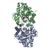 1p6bSC S: Starting model for refinement C: citing same article ( |
|---|---|
| Similar structure data |


-Links
 PDBj
PDBj


- Assembly
| Deposited unit | 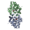
| ||||||||
|---|---|---|---|---|---|---|---|---|---|
| 1 |
| ||||||||
| Unit cell |
|
-Components
| #1: Protein | Mass: 36330.312 Da / Num. of mol.: 2 / Mutation: H254G, H257W, L303T Source method: isolated from a genetically manipulated source Source: (gene. exp.) Flavobacterium sp. (bacteria) / Gene: OPD / Production host: #2: Chemical | ChemComp-ZN /   Mass: 65.409 Da / Num. of mol.: 4 / Source method: obtained synthetically / Formula: Zn Mass: 65.409 Da / Num. of mol.: 4 / Source method: obtained synthetically / Formula: Zn#3: Chemical |   Mass: 242.251 Da / Num. of mol.: 2 / Source method: obtained synthetically / Formula: C12H19O3P Mass: 242.251 Da / Num. of mol.: 2 / Source method: obtained synthetically / Formula: C12H19O3P#4: Chemical |   Mass: 180.182 Da / Num. of mol.: 2 / Source method: obtained synthetically / Formula: C7H17O3P Mass: 180.182 Da / Num. of mol.: 2 / Source method: obtained synthetically / Formula: C7H17O3P#5: Water | ChemComp-HOH / |  Mass: 18.015 Da / Num. of mol.: 321 / Source method: isolated from a natural source / Formula: H2O Mass: 18.015 Da / Num. of mol.: 321 / Source method: isolated from a natural source / Formula: H2O |
|---|
-Experimental details
-Experiment
| Experiment | Method: X-RAY DIFFRACTION / Number of used crystals: 1 |
|---|
- Sample preparation
| Crystal | Density Matthews: 2.75 Å3/Da / Density % sol: 54.86 % |
|---|---|
| Crystal grow | Temperature: 277 K / Method: batch / pH: 9 Details: PEG8000, CHES, NaCl, diethyl 4-methylbenzylphosphonate, diisopropylmethylphosphonate, pH 9, batch, temperature 277K |
| Crystal grow | *PLUS Method: unknown |
-Data collection
| Diffraction | Mean temperature: 277 K |
|---|---|
| Diffraction source | Source: ROTATING ANODE / Type: RIGAKU RU200 / Wavelength: 1.5418 Å |
| Detector | Type: SIEMENS HI-STAR / Detector: AREA DETECTOR / Date: Aug 10, 2001 / Details: supper long mirrors |
| Radiation | Monochromator: Ni Filter / Protocol: SINGLE WAVELENGTH / Monochromatic (M) / Laue (L): M / Scattering type: x-ray |
| Radiation wavelength | Wavelength: 1.5418 Å / Relative weight: 1 |
| Reflection | Resolution: 2→30 Å / Num. all: 47394 / Num. obs: 47394 / % possible obs: 85.3 % / Observed criterion σ(F): 0 / Observed criterion σ(I): 0 / Redundancy: 1.9 % / Rsym value: 0.088 / Net I/σ(I): 8 |
| Reflection shell | Resolution: 2→2.09 Å / Redundancy: 1.4 % / Mean I/σ(I) obs: 1.5 / Num. unique all: 4850 / Rsym value: 0.328 / % possible all: 66.7 |
| Reflection | *PLUS |
- Processing
| Software |
| |||||||||||||||||||||||||
|---|---|---|---|---|---|---|---|---|---|---|---|---|---|---|---|---|---|---|---|---|---|---|---|---|---|---|
| Refinement | Method to determine structure: FOURIER SYNTHESIS Starting model: pdb entry 1P6B Resolution: 2→30 Å / Cross valid method: THROUGHOUT / σ(F): 0 / Stereochemistry target values: Engh & Huber
| |||||||||||||||||||||||||
| Refinement step | Cycle: LAST / Resolution: 2→30 Å
| |||||||||||||||||||||||||
| Refine LS restraints |
| |||||||||||||||||||||||||
| Refinement | *PLUS | |||||||||||||||||||||||||
| Solvent computation | *PLUS | |||||||||||||||||||||||||
| Displacement parameters | *PLUS |
