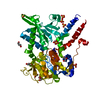
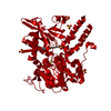
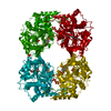
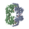
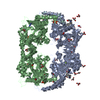
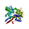
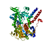
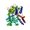
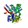
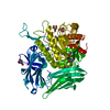
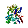
- PDB-6fiw: Human cytosolic 5'-nucleotidase II co-crystallized with 10mM Sodi... -
+
Open data
ID or keywords:
Loading...
-
Basic information
Entry
Database: PDB / ID: 6fiw
Title
Human cytosolic 5'-nucleotidase II co-crystallized with 10mM Sodium ((4-(3'-((7H-purin-6-yl)carbamoyl)-[1,1'-biphenyl]-3-yl)-1H-imidazol-1-yl) methyl) phosphonate
Components
Cytosolic purine 5'-nucleotidase
Keywords
HYDROLASE
Function / homology
Function and homology information
nucleoside phosphotransferase / : / nucleoside phosphotransferase activity / GMP metabolic process / Abacavir metabolism / dGMP metabolic process / negative regulation of defense response to virus by host / : / IMP-specific 5'-nucleotidase / adenosine metabolic process ...nucleoside phosphotransferase / : / nucleoside phosphotransferase activity / GMP metabolic process / Abacavir metabolism / dGMP metabolic process / negative regulation of defense response to virus by host / : / IMP-specific 5'-nucleotidase / adenosine metabolic process / Ribavirin ADME / IMP catabolic process / IMP metabolic process / dGMP catabolic process / allantoin metabolic process / Purine catabolism / 5'-nucleotidase / 5'-nucleotidase activity / protein K48-linked ubiquitination / ubiquitin protein ligase activity / ATP binding / metal ion binding / identical protein binding / cytosol / cytoplasm Similarity search - Function
In the structure databanks used in Yorodumi, some data are registered as the other names, "COVID-19 virus" and "2019-nCoV". Here are the details of the virus and the list of structure data.
Jan 31, 2019. EMDB accession codes are about to change! (news from PDBe EMDB page)
EMDB accession codes are about to change! (news from PDBe EMDB page)
The allocation of 4 digits for EMDB accession codes will soon come to an end. Whilst these codes will remain in use, new EMDB accession codes will include an additional digit and will expand incrementally as the available range of codes is exhausted. The current 4-digit format prefixed with “EMD-” (i.e. EMD-XXXX) will advance to a 5-digit format (i.e. EMD-XXXXX), and so on. It is currently estimated that the 4-digit codes will be depleted around Spring 2019, at which point the 5-digit format will come into force.
The EM Navigator/Yorodumi systems omit the EMD- prefix.
Related info.:Q: What is EMD? / ID/Accession-code notation in Yorodumi/EM Navigator
Yorodumi is a browser for structure data from EMDB, PDB, SASBDB, etc.
This page is also the successor to EM Navigator detail page, and also detail information page/front-end page for Omokage search.
The word "yorodu" (or yorozu) is an old Japanese word meaning "ten thousand". "mi" (miru) is to see.
Related info.:EMDB / PDB / SASBDB / Comparison of 3 databanks / Yorodumi Search / Aug 31, 2016. New EM Navigator & Yorodumi / Yorodumi Papers / Jmol/JSmol / Function and homology information / Changes in new EM Navigator and Yorodumi
 Movie
Movie Controller

 Yorodumi
 Open data
 Basic information
 Components
 Keywords
 Function and homology information
 Homo sapiens (human)
 X-RAY DIFFRACTION /
  Authors
 France, 2items
  Citation
 Structure visualization
 Downloads & links
 Other downloads
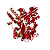


 PDBj
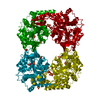
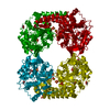
PDBj Assembly
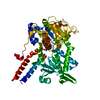


 Mass: 96.063 Da / Num. of mol.: 2 / Source method: obtained synthetically / Formula: SO4
 Mass: 24.305 Da / Num. of mol.: 1 / Source method: obtained synthetically / Formula: Mg
 Mass: 92.094 Da / Num. of mol.: 3 / Source method: obtained synthetically / Formula: C3H8O3
 Mass: 18.015 Da / Num. of mol.: 137 / Source method: isolated from a natural source / Formula: H2O
 Sample preparation
 Processing