 Movie
Movie Controller

[English] 日本語
 Yorodumi
- PDB-6de3: Crystal structure of the double mutant (R39Q/D52N) of the full-le... -
+ Open data

- Basic information
| Entry | Database: PDB / ID: 6de3 | |||||||||||||||
|---|---|---|---|---|---|---|---|---|---|---|---|---|---|---|---|---|
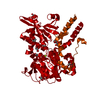
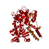
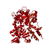
| Title | Crystal structure of the double mutant (R39Q/D52N) of the full-length NT5C2 in the active state | |||||||||||||||
 Components Components | Cytosolic purine 5'-nucleotidase | |||||||||||||||
 Keywords Keywords | HYDROLASE | |||||||||||||||
| Function / homology |  Function and homology information Function and homology informationnucleoside phosphotransferase / : / nucleoside phosphotransferase activity / GMP metabolic process / Abacavir metabolism / dGMP metabolic process / negative regulation of defense response to virus by host / : / IMP-specific 5'-nucleotidase / adenosine metabolic process ...nucleoside phosphotransferase / : / nucleoside phosphotransferase activity / GMP metabolic process / Abacavir metabolism / dGMP metabolic process / negative regulation of defense response to virus by host / : / IMP-specific 5'-nucleotidase / adenosine metabolic process / Ribavirin ADME / IMP catabolic process / IMP metabolic process / dGMP catabolic process / allantoin metabolic process / Purine catabolism / 5'-nucleotidase / 5'-nucleotidase activity / protein K48-linked ubiquitination / ubiquitin protein ligase activity / ATP binding / metal ion binding / identical protein binding / cytosol / cytoplasm Similarity search - Function | |||||||||||||||
| Biological species |  Homo sapiens (human) Homo sapiens (human) | |||||||||||||||
| Method |  X-RAY DIFFRACTION / SYNCHROTRON / MOLECULAR REPLACEMENT / Resolution: 3.06 Å X-RAY DIFFRACTION / SYNCHROTRON / MOLECULAR REPLACEMENT / Resolution: 3.06 Å | |||||||||||||||
 Authors Authors | Forouhar, F. / Dieck, C.L. / Tzoneva, G. / Carpenter, Z. / Ambesi-Impiombato, A. / Sanchez-Martin, M. / Kirschner-Schwabe, R. / Lew, S. / Seetharaman, J. / Ferrando, A.A. / Tong, L. | |||||||||||||||
| Funding support |  United States, 4items United States, 4items
| |||||||||||||||
 Citation Citation | Journal: Cancer Cell / Year: 2018 Title: Structure and Mechanisms of NT5C2 Mutations Driving Thiopurine Resistance in Relapsed Lymphoblastic Leukemia. Authors: Dieck, C.L. / Tzoneva, G. / Forouhar, F. / Carpenter, Z. / Ambesi-Impiombato, A. / Sanchez-Martin, M. / Kirschner-Schwabe, R. / Lew, S. / Seetharaman, J. / Tong, L. / Ferrando, A.A. | |||||||||||||||
| History |
|
- Structure visualization
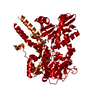
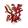
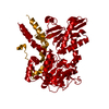
| Structure viewer | Molecule: MolmilJmol/JSmol |
|---|
- Downloads & links
-Download
| PDBx/mmCIF format | 6de3.cif.gz | 208.7 KB | Display | PDBx/mmCIF format |
|---|---|---|---|---|
| PDB format | pdb6de3.ent.gz | 160.9 KB | Display | PDB format |
| PDBx/mmJSON format | 6de3.json.gz | Tree view | PDBx/mmJSON format | |
| Others |  Other downloads Other downloads |
-Validation report
| Arichive directory | https://data.pdbj.org/pub/pdb/validation_reports/de/6de3ftp://data.pdbj.org/pub/pdb/validation_reports/de/6de3 | HTTPS FTP |
|---|
-Related structure data
| Related structure data | 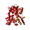 6dd3C 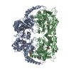 6ddbC 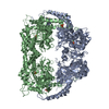 6ddcC 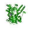 6ddhSC 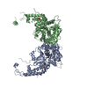 6ddkC 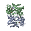 6ddlC 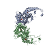 6ddoC 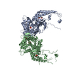 6ddqC 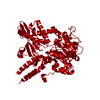 6ddxC 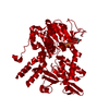 6ddyC  6ddzC 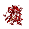 6de0C 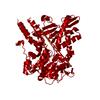 6de1C 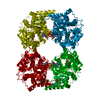 6de2C S: Starting model for refinement C: citing same article ( |
|---|---|
| Similar structure data |


-Links
 PDBj
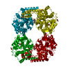
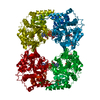
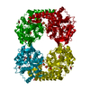
PDBj- Assembly
| Deposited unit | 
| ||||||||
|---|---|---|---|---|---|---|---|---|---|
| 1 | 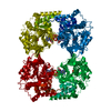
| ||||||||
| Unit cell |
| ||||||||
| Components on special symmetry positions |
|
-Components
| #1: Protein | Mass: 66926.672 Da / Num. of mol.: 1 / Mutation: R39Q, D52N Source method: isolated from a genetically manipulated source Source: (gene. exp.) Homo sapiens (human) / Gene: NT5C2, NT5B, NT5CP, PNT5 / Plasmid: pLOC_NT5C2 / Cell (production host): Rosetta 2(DE3) / Production host:  |
|---|---|
| #2: Chemical | ChemComp-PO4 /   Mass: 94.971 Da / Num. of mol.: 1 / Source method: obtained synthetically / Formula: PO4 Mass: 94.971 Da / Num. of mol.: 1 / Source method: obtained synthetically / Formula: PO4 |
| #3: Chemical | ChemComp-ATP /   Mass: 507.181 Da / Num. of mol.: 1 / Source method: obtained synthetically / Formula: C10H16N5O13P3 / Comment: ATP, energy-carrying molecule*YM Mass: 507.181 Da / Num. of mol.: 1 / Source method: obtained synthetically / Formula: C10H16N5O13P3 / Comment: ATP, energy-carrying molecule*YM |
| #4: Chemical | ChemComp-MG /   Mass: 24.305 Da / Num. of mol.: 1 / Source method: obtained synthetically / Formula: Mg Mass: 24.305 Da / Num. of mol.: 1 / Source method: obtained synthetically / Formula: Mg |
| #5: Water | ChemComp-HOH /  Mass: 18.015 Da / Num. of mol.: 50 / Source method: isolated from a natural source / Formula: H2O Mass: 18.015 Da / Num. of mol.: 50 / Source method: isolated from a natural source / Formula: H2O |
-Experimental details
-Experiment
| Experiment | Method: X-RAY DIFFRACTION / Number of used crystals: 1 |
|---|
- Sample preparation
| Crystal | Density Matthews: 3 Å3/Da / Density % sol: 57 % |
|---|---|
| Crystal grow | Temperature: 293 K / Method: microbatch / pH: 7.5 Details: 100 mM HEPES (pH 7.5), 12% (w/v) PEG 3350, 5 mM ATP, 5 mM IMP, and 5 mM MgCl2 |
-Data collection
| Diffraction | Mean temperature: 100 K |
|---|---|
| Diffraction source | Source: SYNCHROTRON / Site: APS / Beamline: 19-ID / Wavelength: 0.979 Å |
| Detector | Type: SBC-2 / Detector: CCD / Date: Oct 29, 2015 |
| Radiation | Protocol: SINGLE WAVELENGTH / Monochromatic (M) / Laue (L): M / Scattering type: x-ray |
| Radiation wavelength | Wavelength: 0.979 Å / Relative weight: 1 |
| Reflection | Resolution: 3.06→46.181 Å / Num. obs: 14798 / % possible obs: 99.2 % / Observed criterion σ(F): 0 / Observed criterion σ(I): 0 / Redundancy: 4.7 % / Biso Wilson estimate: 50.18 Å2 / CC1/2: 0.96 / Rmerge(I) obs: 0.14 / Rpim(I) all: 0.07 / Rrim(I) all: 0.15 / Χ2: 0.96 / Net I/av σ(I): 9.8 / Net I/σ(I): 9.8 |
| Reflection shell | Resolution: 3.06→3.12 Å / Redundancy: 4.7 % / Rmerge(I) obs: 0.56 / Mean I/σ(I) obs: 2.9 / Num. unique obs: 872 / CC1/2: 0.86 / Rpim(I) all: 0.25 / Rrim(I) all: 0.56 / Χ2: 0.84 / % possible all: 99.9 |
- Processing
| Software |
| ||||||||||||||||||||||||||||||||||||||||||||||||||||||||||||||||||||||||||||||||||||
|---|---|---|---|---|---|---|---|---|---|---|---|---|---|---|---|---|---|---|---|---|---|---|---|---|---|---|---|---|---|---|---|---|---|---|---|---|---|---|---|---|---|---|---|---|---|---|---|---|---|---|---|---|---|---|---|---|---|---|---|---|---|---|---|---|---|---|---|---|---|---|---|---|---|---|---|---|---|---|---|---|---|---|---|---|---|
| Refinement | Method to determine structure: MOLECULAR REPLACEMENT Starting model: 6DDH Resolution: 3.06→46.181 Å / SU ML: 0.39 / Cross valid method: FREE R-VALUE / σ(F): 1.35 / Phase error: 23.39
| ||||||||||||||||||||||||||||||||||||||||||||||||||||||||||||||||||||||||||||||||||||
| Solvent computation | Shrinkage radii: 0.9 Å / VDW probe radii: 1.11 Å | ||||||||||||||||||||||||||||||||||||||||||||||||||||||||||||||||||||||||||||||||||||
| Refinement step | Cycle: LAST / Resolution: 3.06→46.181 Å
| ||||||||||||||||||||||||||||||||||||||||||||||||||||||||||||||||||||||||||||||||||||
| Refine LS restraints |
| ||||||||||||||||||||||||||||||||||||||||||||||||||||||||||||||||||||||||||||||||||||
| LS refinement shell |
| ||||||||||||||||||||||||||||||||||||||||||||||||||||||||||||||||||||||||||||||||||||
| Refinement TLS params. | Method: refined / Origin x: -0.1124 Å / Origin y: 39.3518 Å / Origin z: 19.2402 Å
| ||||||||||||||||||||||||||||||||||||||||||||||||||||||||||||||||||||||||||||||||||||
| Refinement TLS group | Selection details: all |
