 Movie
Movie Controller

[English] 日本語
 Yorodumi
- PDB-6fgr: Crystal Structure of the Amyloid-like IIKIIK Segment from the S. ... -
+ Open data

- Basic information
| Entry | Database: PDB / ID: 6fgr | |||||||||||||||
|---|---|---|---|---|---|---|---|---|---|---|---|---|---|---|---|---|
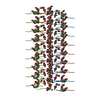
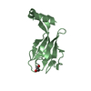
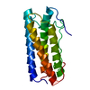
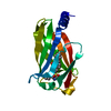
| Title | Crystal Structure of the Amyloid-like IIKIIK Segment from the S. aureus Biofilm-associated PSMalpha4 | |||||||||||||||
 Components Components | Psm alpha-4 | |||||||||||||||
 Keywords Keywords | PROTEIN FIBRIL / steric-zipper / cross-beta / bacterial amyloid fibril / S. aureus / PSM | |||||||||||||||
| Function / homology | Phenol-soluble modulin alpha peptide / Phenol-soluble modulin alpha peptide family / killing of cells of another organism / Phenol-soluble modulin PSM-alpha-4 Function and homology information Function and homology information | |||||||||||||||
| Biological species |   Staphylococcus aureus (bacteria) Staphylococcus aureus (bacteria) | |||||||||||||||
| Method |  X-RAY DIFFRACTION / SYNCHROTRON / MOLECULAR REPLACEMENT / molecular replacement / Resolution: 1.5 Å X-RAY DIFFRACTION / SYNCHROTRON / MOLECULAR REPLACEMENT / molecular replacement / Resolution: 1.5 Å | |||||||||||||||
| Model details | Phenol Soluble Modulin | |||||||||||||||
 Authors Authors | Landau, M. / Colletier, J.-P. | |||||||||||||||
| Funding support |  Israel, 4items Israel, 4items
| |||||||||||||||
 Citation Citation | Journal: Nat Commun / Year: 2018 Title: Extreme amyloid polymorphism in Staphylococcus aureus virulent PSM alpha peptides. Authors: Salinas, N. / Colletier, J.P. / Moshe, A. / Landau, M. | |||||||||||||||
| History |
|
- Structure visualization
| Structure viewer | Molecule: MolmilJmol/JSmol |
|---|
- Downloads & links
-Download
| PDBx/mmCIF format | 6fgr.cif.gz | 15 KB | Display | PDBx/mmCIF format |
|---|---|---|---|---|
| PDB format | pdb6fgr.ent.gz | 8.9 KB | Display | PDB format |
| PDBx/mmJSON format | 6fgr.json.gz | Tree view | PDBx/mmJSON format | |
| Others |  Other downloads Other downloads |
-Validation report
| Arichive directory | https://data.pdbj.org/pub/pdb/validation_reports/fg/6fgrftp://data.pdbj.org/pub/pdb/validation_reports/fg/6fgr | HTTPS FTP |
|---|
-Related structure data


-Links
 PDBj
PDBj- Assembly
| Deposited unit | 
| ||||||||
|---|---|---|---|---|---|---|---|---|---|
| 1 | 
| ||||||||
| Unit cell |
|
-Components
| #1: Protein/peptide | Mass: 729.006 Da / Num. of mol.: 2 Fragment: Amyloid spine segment IIKIIK from PSMalpha4 (residues 7-12) secreted by S. aureus Source method: obtained synthetically / Details: IIKIIK from PSMalpha4, synthesized / Source: (synth.) Staphylococcus aureus (bacteria) / References: UniProt: H9BRQ8#2: Chemical | ChemComp-EDO / |   Mass: 62.068 Da / Num. of mol.: 1 / Source method: obtained synthetically / Formula: C2H6O2 Mass: 62.068 Da / Num. of mol.: 1 / Source method: obtained synthetically / Formula: C2H6O2#3: Chemical |   Mass: 96.063 Da / Num. of mol.: 2 / Source method: obtained synthetically / Formula: SO4 Mass: 96.063 Da / Num. of mol.: 2 / Source method: obtained synthetically / Formula: SO4#4: Water | ChemComp-HOH / |  Mass: 18.015 Da / Num. of mol.: 3 / Source method: isolated from a natural source / Formula: H2O Mass: 18.015 Da / Num. of mol.: 3 / Source method: isolated from a natural source / Formula: H2O |
|---|
-Experimental details
-Experiment
| Experiment | Method: X-RAY DIFFRACTION / Number of used crystals: 1 |
|---|
- Sample preparation
| Crystal | Density Matthews: 1.62 Å3/Da / Density % sol: 24.28 % |
|---|---|
| Crystal grow | Temperature: 293 K / Method: vapor diffusion, hanging drop Details: Reservoir contained 0.2M Ammonium sulfate, 20% polyethylene glycol 3350 |
-Data collection
| Diffraction | Mean temperature: 100 K | ||||||||||||||||||||||||||||||||||||||||||||||||||||||||||||||||||||||||||||||||||||||||||||||||||||||||||||||||||||||||||||||||||||||||||||||||||||||
|---|---|---|---|---|---|---|---|---|---|---|---|---|---|---|---|---|---|---|---|---|---|---|---|---|---|---|---|---|---|---|---|---|---|---|---|---|---|---|---|---|---|---|---|---|---|---|---|---|---|---|---|---|---|---|---|---|---|---|---|---|---|---|---|---|---|---|---|---|---|---|---|---|---|---|---|---|---|---|---|---|---|---|---|---|---|---|---|---|---|---|---|---|---|---|---|---|---|---|---|---|---|---|---|---|---|---|---|---|---|---|---|---|---|---|---|---|---|---|---|---|---|---|---|---|---|---|---|---|---|---|---|---|---|---|---|---|---|---|---|---|---|---|---|---|---|---|---|---|---|---|---|
| Diffraction source | Source: SYNCHROTRON / Site: ESRF  / Beamline: ID23-2 / Wavelength: 0.8729 Å / Beamline: ID23-2 / Wavelength: 0.8729 Å | ||||||||||||||||||||||||||||||||||||||||||||||||||||||||||||||||||||||||||||||||||||||||||||||||||||||||||||||||||||||||||||||||||||||||||||||||||||||
| Detector | Type: DECTRIS PILATUS 2M / Detector: PIXEL / Date: Oct 8, 2014 | ||||||||||||||||||||||||||||||||||||||||||||||||||||||||||||||||||||||||||||||||||||||||||||||||||||||||||||||||||||||||||||||||||||||||||||||||||||||
| Radiation | Protocol: SINGLE WAVELENGTH / Monochromatic (M) / Laue (L): M / Scattering type: x-ray | ||||||||||||||||||||||||||||||||||||||||||||||||||||||||||||||||||||||||||||||||||||||||||||||||||||||||||||||||||||||||||||||||||||||||||||||||||||||
| Radiation wavelength | Wavelength: 0.8729 Å / Relative weight: 1 | ||||||||||||||||||||||||||||||||||||||||||||||||||||||||||||||||||||||||||||||||||||||||||||||||||||||||||||||||||||||||||||||||||||||||||||||||||||||
| Reflection | Resolution: 1.5→18.21 Å / Num. obs: 1397 / % possible obs: 95 % / Redundancy: 9.89 % / Biso Wilson estimate: 16.205 Å2 / CC1/2: 0.994 / Rmerge(I) obs: 0.206 / Rrim(I) all: 0.217 / Χ2: 0.835 / Net I/σ(I): 7.18 / Num. measured all: 13816 / Scaling rejects: 1 | ||||||||||||||||||||||||||||||||||||||||||||||||||||||||||||||||||||||||||||||||||||||||||||||||||||||||||||||||||||||||||||||||||||||||||||||||||||||
| Reflection shell | Diffraction-ID: 1
|
-Phasing
| Phasing | Method: molecular replacement |
|---|---|
| Phasing MR | Model details: Phaser MODE: MR_AUTO / Packing: 0 |
- Processing
| Software |
| |||||||||||||||||||||||||||||||||||||||||||||||||||||||||||||||||
|---|---|---|---|---|---|---|---|---|---|---|---|---|---|---|---|---|---|---|---|---|---|---|---|---|---|---|---|---|---|---|---|---|---|---|---|---|---|---|---|---|---|---|---|---|---|---|---|---|---|---|---|---|---|---|---|---|---|---|---|---|---|---|---|---|---|---|
| Refinement | Method to determine structure: MOLECULAR REPLACEMENT Starting model: ideal beta-strand of poly-ala Resolution: 1.5→18.21 Å / Cor.coef. Fo:Fc: 0.959 / Cor.coef. Fo:Fc free: 0.944 / WRfactor Rfree: 0.2222 / WRfactor Rwork: 0.1868 / FOM work R set: 0.8212 / SU B: 4.347 / SU ML: 0.071 / SU R Cruickshank DPI: 0.3052 / SU Rfree: 0.1113 / Cross valid method: THROUGHOUT / σ(F): 0 / ESU R: 0.305 / ESU R Free: 0.111 / Stereochemistry target values: MAXIMUM LIKELIHOOD Details: HYDROGENS HAVE BEEN ADDED IN THE RIDING POSITIONS U VALUES : REFINED INDIVIDUALLY
| |||||||||||||||||||||||||||||||||||||||||||||||||||||||||||||||||
| Solvent computation | Ion probe radii: 0.8 Å / Shrinkage radii: 0.8 Å / VDW probe radii: 1.2 Å / Solvent model: MASK | |||||||||||||||||||||||||||||||||||||||||||||||||||||||||||||||||
| Displacement parameters | Biso max: 61.86 Å2 / Biso mean: 9.128 Å2 / Biso min: 1.71 Å2
| |||||||||||||||||||||||||||||||||||||||||||||||||||||||||||||||||
| Refinement step | Cycle: final / Resolution: 1.5→18.21 Å
| |||||||||||||||||||||||||||||||||||||||||||||||||||||||||||||||||
| Refine LS restraints |
| |||||||||||||||||||||||||||||||||||||||||||||||||||||||||||||||||
| LS refinement shell | Resolution: 1.502→1.54 Å / Rfactor Rfree error: 0 / Total num. of bins used: 20
|
