 Movie
Movie Controller


+ Open data

- Basic information
| Entry | Database: PDB / ID: 2w2s | ||||||
|---|---|---|---|---|---|---|---|
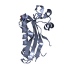
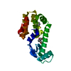
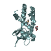
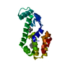
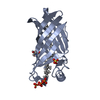
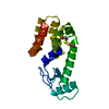
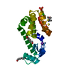
| Title | Structure of the Lagos bat virus matrix protein | ||||||
 Components Components | MATRIX PROTEIN | ||||||
 Keywords Keywords | VIRAL PROTEIN / VIRAL ASSEMBLY / VIRAL MORPHOGENESIS / LAGOS BAT VIRUS / POLYMER / MATRIX PROTEIN / VSV | ||||||
| Function / homology |  Function and homology information Function and homology informationhost cell endomembrane system / viral budding via host ESCRT complex / structural constituent of virion / viral envelope / virion membrane Similarity search - Function | ||||||
| Biological species |  LAGOS BAT VIRUS LAGOS BAT VIRUS | ||||||
| Method |  X-RAY DIFFRACTION / SYNCHROTRON / MAD / Resolution: 2.75 Å X-RAY DIFFRACTION / SYNCHROTRON / MAD / Resolution: 2.75 Å | ||||||
 Authors Authors | Graham, S.C. / Assenberg, R. / Delmas, O. / Verma, A. / Gholami, A. / Talbi, C. / Owens, R.J. / Stuart, D.I. / Grimes, J.M. / Bourhy, H. | ||||||
 Citation Citation | Journal: Plos Pathog. / Year: 2008 Title: Rhabdovirus Matrix Protein Structures Reveal a Novel Mode of Self-Association. Authors: Graham, S.C. / Assenberg, R. / Delmas, O. / Verma, A. / Gholami, A. / Talbi, C. / Owens, R.J. / Stuart, D.I. / Grimes, J.M. / Bourhy, H. #1: Journal: Acta Crystallogr.,Sect.F / Year: 2008 Title: Expression, Purification and Crystallization of a Lyssavirus Matrix (M) Protein. Authors: Assenberg, R. / Delmas, O. / Graham, S.C. / Verma, A. / Berrow, N. / Stuart, D.I. / Owens, R.J. / Bourhy, H. / Grimes, J.M. | ||||||
| History |
| ||||||
| Remark 700 | SHEET THE SHEET STRUCTURE OF THIS MOLECULE IS BIFURCATED. IN ORDER TO REPRESENT THIS FEATURE IN ... SHEET THE SHEET STRUCTURE OF THIS MOLECULE IS BIFURCATED. IN ORDER TO REPRESENT THIS FEATURE IN THE SHEET RECORDS BELOW, TWO SHEETS ARE DEFINED. |
- Structure visualization
| Structure viewer | Molecule: MolmilJmol/JSmol |
|---|
- Downloads & links
-Download
| PDBx/mmCIF format | 2w2s.cif.gz | 80.4 KB | Display | PDBx/mmCIF format |
|---|---|---|---|---|
| PDB format | pdb2w2s.ent.gz | 61.8 KB | Display | PDB format |
| PDBx/mmJSON format | 2w2s.json.gz | Tree view | PDBx/mmJSON format | |
| Others |  Other downloads Other downloads |
-Validation report
| Arichive directory | https://data.pdbj.org/pub/pdb/validation_reports/w2/2w2sftp://data.pdbj.org/pub/pdb/validation_reports/w2/2w2s | HTTPS FTP |
|---|
-Related structure data


-Links
 PDBj
PDBj- Assembly
| Deposited unit | 
| ||||||||
|---|---|---|---|---|---|---|---|---|---|
| 1 |
| ||||||||
| Unit cell |
| ||||||||
| Details | THE PROTEIN IS A NON-COVALENT LINEAR POLYMER WHERE GLOBULAR DOMAINS (RESIDUES ARE 48-202) ARE NON-COVALENTLY ASSOCIATED BY A FLEXIBLE LINKER, WITH RESIDUES 30-37 MEDIATING THE INTER-MOLECULAR INTERACTION. RESIDUES 30-37, WHICH INTERACT WITH THE GLOBULAR DOMAIN (RESIDUES 48-202) IN THE LOOPS BETWEEN BETA SHEET 1 TO ALPHA HELIX 1 AND BETA SHEET 2 TO BETA SHEET 3, ARE NOT COVALENTLY LINKED TO THIS GLOBULAR DOMAIN. RATHER, THEY ARE COVALENTLY LINKED TO AN ADJACENT GLOBULAR DOMAIN IN THE CRYSTAL RELATED BY THE SYMMETRY OPERATOR [1+X-Y,1-Y,1-Z]. REPEATED, THIS INTER-MOLECULAR INTERACTION GIVES RISE TO LINEAR POLYMERS OF THE M PROTEIN WHERE MOLECULES ARE NON-COVALENTLY LINKED VIA THE INTERACTION BETWEEN RESIDUES 30-37 AND THE GLOBULAR DOMAIN. IN ORDER TO GENERATE THE LINEAR POLYMER THE FOLLOWING TRANSFORMATION MATRIX SHOULD BE APPLIED: RX RY RZ T 1.0000 0.0000 0.0000 28.4350 0.0000 -1.0000 0.0000 49.2510 0.0000 0.0000 -1.0000 187.9100 |
-Components
| #1: Protein | Mass: 23177.535 Da / Num. of mol.: 1 Source method: isolated from a genetically manipulated source Source: (gene. exp.) LAGOS BAT VIRUSDescription: ISOLATE 8619NGA, GENOTYPE 2, ISOLATED FROM A FRUGIVOROUS BAT IN NIGERIA (BOULGER, L. R., AND J. S. PORTEFIELD. 1958. ISOLATION OF A VIRUS FROM NIGERIAN FRUIT BATS. TRANS.R.SOC.TROP.MED.HYG. 52\:421-424.) Plasmid: POPINS / Production host:  |
|---|---|
| Has protein modification | Y |
-Experimental details
-Experiment
| Experiment | Method: X-RAY DIFFRACTION / Number of used crystals: 1 |
|---|
- Sample preparation
| Crystal | Density Matthews: 1.91 Å3/Da / Density % sol: 35.61 % / Description: NONE |
|---|---|
| Crystal grow | Method: vapor diffusion, sitting drop / pH: 4 Details: SITTING DROPS CONTAINING 100 NL OF 1.1 MG/ML PROTEIN AND 100 NL OF RESERVOIR SOLUTION (100 MM CITRATE PH 4.0 AND 10%(W/V) POLYETHYLENE GLYCOL (PEG) 6000) WERE EQUILIBRATED AGAINST 95 UL ...Details: SITTING DROPS CONTAINING 100 NL OF 1.1 MG/ML PROTEIN AND 100 NL OF RESERVOIR SOLUTION (100 MM CITRATE PH 4.0 AND 10%(W/V) POLYETHYLENE GLYCOL (PEG) 6000) WERE EQUILIBRATED AGAINST 95 UL RESERVOIRS AT 20.5C. CRYSTALS WERE CRYOPROTECTED BY BRIEF IMMERSION IN RESERVOIR SOLUTION SUPPLEMENTED WITH 25% V/V GLYCEROL. |
-Data collection
| Diffraction | Mean temperature: 100 K |
|---|---|
| Diffraction source | Source: SYNCHROTRON / Site: ESRF  / Beamline: ID23-2 / Wavelength: 0.8726 / Beamline: ID23-2 / Wavelength: 0.8726 |
| Detector | Type: MARRESEARCH / Detector: CCD / Date: Nov 4, 2006 / Details: KB PAIR PT COATED SI MIRRORS |
| Radiation | Monochromator: SI (111) CRYSTAL / Protocol: MAD / Monochromatic (M) / Laue (L): M / Scattering type: x-ray |
| Radiation wavelength | Wavelength: 0.8726 Å / Relative weight: 1 |
| Reflection | Resolution: 2.75→38.72 Å / Num. obs: 5164 / % possible obs: 99.6 % / Observed criterion σ(I): -3 / Redundancy: 6.2 % / Biso Wilson estimate: 59.7 Å2 / Rmerge(I) obs: 0.13 / Net I/σ(I): 10.2 |
| Reflection shell | Resolution: 2.75→2.82 Å / Redundancy: 5.4 % / Rmerge(I) obs: 0.77 / Mean I/σ(I) obs: 2.2 / % possible all: 99.4 |
- Processing
| Software |
| ||||||||||||||||||||||||||||||||||||||||||||||||||||||||||||||||||||||||||||||||||||||||||||||||||||||||||||||||||||||||||||||||||||||||||||||||||||||||||||||||||||||||||||||||||||||
|---|---|---|---|---|---|---|---|---|---|---|---|---|---|---|---|---|---|---|---|---|---|---|---|---|---|---|---|---|---|---|---|---|---|---|---|---|---|---|---|---|---|---|---|---|---|---|---|---|---|---|---|---|---|---|---|---|---|---|---|---|---|---|---|---|---|---|---|---|---|---|---|---|---|---|---|---|---|---|---|---|---|---|---|---|---|---|---|---|---|---|---|---|---|---|---|---|---|---|---|---|---|---|---|---|---|---|---|---|---|---|---|---|---|---|---|---|---|---|---|---|---|---|---|---|---|---|---|---|---|---|---|---|---|---|---|---|---|---|---|---|---|---|---|---|---|---|---|---|---|---|---|---|---|---|---|---|---|---|---|---|---|---|---|---|---|---|---|---|---|---|---|---|---|---|---|---|---|---|---|---|---|---|---|
| Refinement | Method to determine structure: MAD Starting model: NONE Resolution: 2.75→38.72 Å / Cor.coef. Fo:Fc: 0.934 / Cor.coef. Fo:Fc free: 0.893 / SU B: 29.934 / SU ML: 0.269 / Cross valid method: THROUGHOUT / ESU R Free: 0.374 / Stereochemistry target values: MAXIMUM LIKELIHOOD / Details: HYDROGENS HAVE BEEN ADDED IN THE RIDING POSITIONS.
| ||||||||||||||||||||||||||||||||||||||||||||||||||||||||||||||||||||||||||||||||||||||||||||||||||||||||||||||||||||||||||||||||||||||||||||||||||||||||||||||||||||||||||||||||||||||
| Solvent computation | Ion probe radii: 0.8 Å / Shrinkage radii: 0.8 Å / VDW probe radii: 1.2 Å / Solvent model: MASK | ||||||||||||||||||||||||||||||||||||||||||||||||||||||||||||||||||||||||||||||||||||||||||||||||||||||||||||||||||||||||||||||||||||||||||||||||||||||||||||||||||||||||||||||||||||||
| Displacement parameters | Biso mean: 95.8 Å2
| ||||||||||||||||||||||||||||||||||||||||||||||||||||||||||||||||||||||||||||||||||||||||||||||||||||||||||||||||||||||||||||||||||||||||||||||||||||||||||||||||||||||||||||||||||||||
| Refinement step | Cycle: LAST / Resolution: 2.75→38.72 Å
| ||||||||||||||||||||||||||||||||||||||||||||||||||||||||||||||||||||||||||||||||||||||||||||||||||||||||||||||||||||||||||||||||||||||||||||||||||||||||||||||||||||||||||||||||||||||
| Refine LS restraints |
|