 Movie
Movie Controller


+ Open data

- Basic information
| Entry | Database: PDB / ID: 2w2r | ||||||
|---|---|---|---|---|---|---|---|
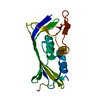
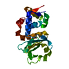
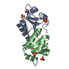
| Title | Structure of the vesicular stomatitis virus matrix protein | ||||||
 Components Components | MATRIX PROTEIN | ||||||
 Keywords Keywords | VIRAL PROTEIN / VIRAL ASSEMBLY / VIRAL MORPHOGENESIS / VSV / POLYMER / MATRIX PROTEIN | ||||||
| Function / homology |  Function and homology information Function and homology informationhost cell nuclear membrane / viral budding via host ESCRT complex / symbiont-mediated suppression of host mRNA export from nucleus / structural constituent of virion / host cell cytoplasm / viral envelope Similarity search - Function | ||||||
| Biological species |  VESICULAR STOMATITIS VIRUS VESICULAR STOMATITIS VIRUS | ||||||
| Method |  X-RAY DIFFRACTION / SYNCHROTRON / SAD / Resolution: 1.83 Å X-RAY DIFFRACTION / SYNCHROTRON / SAD / Resolution: 1.83 Å | ||||||
 Authors Authors | Graham, S.C. / Assenberg, R. / Delmas, O. / Verma, A. / Gholami, A. / Talbi, C. / Owens, R.J. / Stuart, D.I. / Grimes, J.M. / Bourhy, H. | ||||||
 Citation Citation | Journal: Plos Pathog. / Year: 2008 Title: Rhabdovirus Matrix Protein Structures Reveal a Novel Mode of Self-Association. Authors: Graham, S.C. / Assenberg, R. / Delmas, O. / Verma, A. / Gholami, A. / Talbi, C. / Owens, R.J. / Stuart, D.I. / Grimes, J.M. / Bourhy, H. | ||||||
| History |
|
- Structure visualization
| Structure viewer | Molecule: MolmilJmol/JSmol |
|---|
- Downloads & links
-Download
| PDBx/mmCIF format | 2w2r.cif.gz | 91.5 KB | Display | PDBx/mmCIF format |
|---|---|---|---|---|
| PDB format | pdb2w2r.ent.gz | 70 KB | Display | PDB format |
| PDBx/mmJSON format | 2w2r.json.gz | Tree view | PDBx/mmJSON format | |
| Others |  Other downloads Other downloads |
-Validation report
| Arichive directory | https://data.pdbj.org/pub/pdb/validation_reports/w2/2w2rftp://data.pdbj.org/pub/pdb/validation_reports/w2/2w2r | HTTPS FTP |
|---|
-Related structure data


-Links
 PDBj
PDBj- Assembly
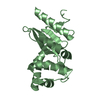
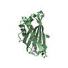
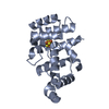
| Deposited unit | 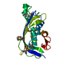
| ||||||||
|---|---|---|---|---|---|---|---|---|---|
| 1 |
| ||||||||
| Unit cell |
| ||||||||
| Details | THE PROTEIN IS A NON-COVALENT LINEAR POLYMER WHERE GLOBULAR DOMAINS (RESIDUES ARE 58-229) ARE NON-COVALENTLY ASSOCIATED BY A FLEXIBLE LINKER, WITH RESIDUES 41-52 MEDIATING THE INTER-MOLECULAR INTERACTION. RESIDUES 41-52, WHICH INTERACT WITH THE GLOBULAR DOMAIN (RESIDUES 58-229) IN THE LOOPS BETWEEN BETA SHEET 1 TO ALPHA HELIX 1, ALPHA HELIX 2 TO ALPHA HELIX 2.5 AND THE LOOP PRECEDING ALPHA HELIX 3, ARE NOT COVALENTLY LINKED TO THIS GLOBULAR DOMAIN. RATHER, THEY ARE COVALENTLY LINKED TO AN ADJACENT GLOBULAR DOMAIN IN THE CRYSTAL RELATED BY THE SYMMETRY OPERATOR [-X-1/2,-Y-1/2,Z-1/2]. REPEATED, THIS INTER-MOLECULAR INTERACTION GIVES RISE TO LINEAR POLYMERS OF THE M PROTEIN WHERE MOLECULES ARE NON-COVALENTLY LINKED VIA THE INTERACTION BETWEEN RESIDUES 41-52 AND THE GLOBULAR DOMAIN. IN ORDER TO GENERATE THE LINEAR POLYMER THE FOLLOWING TRANSFORMATION MATRIX SHOULD BE APPLIED: RX RY RZ T -1.0000 0.0000 0.0000 -43.1300 -0.0000 -1.0000 -0.0000 -43.1300 -0.0000 -0.0000 1.0000 -35.1600 |
-Components
| #1: Protein | Mass: 26525.031 Da / Num. of mol.: 1 Source method: isolated from a genetically manipulated source Source: (gene. exp.) VESICULAR STOMATITIS VIRUS / Strain: NEW JERSEY / Plasmid: POPINS / Production host:  |
|---|---|
| #2: Water | ChemComp-HOH /  Mass: 18.015 Da / Num. of mol.: 147 / Source method: isolated from a natural source / Formula: H2O Mass: 18.015 Da / Num. of mol.: 147 / Source method: isolated from a natural source / Formula: H2O |
| Has protein modification | Y |
| Sequence details | THE SEQUENCE OF THE PROTEIN WAS DERIVED FROM GENBANK ENTRY EU917223 WHICH WAS UNRELEASED AT THE ...THE SEQUENCE OF THE PROTEIN WAS DERIVED FROM GENBANK ENTRY EU917223 WHICH WAS UNRELEASED |
-Experimental details
-Experiment
| Experiment | Method: X-RAY DIFFRACTION / Number of used crystals: 1 |
|---|
- Sample preparation
| Crystal | Density Matthews: 2.52 Å3/Da / Density % sol: 51.13 % / Description: NONE |
|---|---|
| Crystal grow | Method: vapor diffusion, sitting drop / pH: 5.6 Details: SITTING DROPS CONTAINING 100 NL 1.2 MG/ML PROTEIN AND 100 NL OF RESERVOIR SOLUTION (20% V/V ISOPROPANOL, 20% W/V PEG 4000 AND 0.1 M SODIUM CITRATE (PH 5.6)) WERE EQUILIBRATED AGAINST 95 UL ...Details: SITTING DROPS CONTAINING 100 NL 1.2 MG/ML PROTEIN AND 100 NL OF RESERVOIR SOLUTION (20% V/V ISOPROPANOL, 20% W/V PEG 4000 AND 0.1 M SODIUM CITRATE (PH 5.6)) WERE EQUILIBRATED AGAINST 95 UL RESERVIOURS AT 20.5C. CRYSTALS WERE CRYOPROTECTED BY A QUICK SWEEP THROUGH RESERVOIR SUPPLEMENTED WITH 20% V/V GLYCEROL. |
-Data collection
| Diffraction | Mean temperature: 100 K |
|---|---|
| Diffraction source | Source: SYNCHROTRON / Site: ESRF  / Beamline: ID23-1 / Wavelength: 0.9802 / Beamline: ID23-1 / Wavelength: 0.9802 |
| Detector | Type: ADSC CCD / Detector: CCD / Date: Mar 17, 2008 / Details: MIRRORS |
| Radiation | Monochromator: SI (111) CRYSTAL / Protocol: SINGLE WAVELENGTH / Monochromatic (M) / Laue (L): M / Scattering type: x-ray |
| Radiation wavelength | Wavelength: 0.9802 Å / Relative weight: 1 |
| Reflection | Resolution: 1.83→35.16 Å / Num. obs: 22813 / % possible obs: 99.8 % / Observed criterion σ(I): -3 / Redundancy: 7.3 % / Biso Wilson estimate: 21.1 Å2 / Rmerge(I) obs: 0.12 / Net I/σ(I): 10.7 |
| Reflection shell | Resolution: 1.83→1.88 Å / Redundancy: 6.1 % / Rmerge(I) obs: 0.84 / Mean I/σ(I) obs: 2 / % possible all: 99.6 |
- Processing
| Software |
| ||||||||||||||||||||||||||||||||||||||||||||||||||||||||||||||||||||||||||||||||||||||||||||||||||||||||||||||||||||||||||||||||||||||||||||||||||||||||||||||||||||||||||||||||||||||
|---|---|---|---|---|---|---|---|---|---|---|---|---|---|---|---|---|---|---|---|---|---|---|---|---|---|---|---|---|---|---|---|---|---|---|---|---|---|---|---|---|---|---|---|---|---|---|---|---|---|---|---|---|---|---|---|---|---|---|---|---|---|---|---|---|---|---|---|---|---|---|---|---|---|---|---|---|---|---|---|---|---|---|---|---|---|---|---|---|---|---|---|---|---|---|---|---|---|---|---|---|---|---|---|---|---|---|---|---|---|---|---|---|---|---|---|---|---|---|---|---|---|---|---|---|---|---|---|---|---|---|---|---|---|---|---|---|---|---|---|---|---|---|---|---|---|---|---|---|---|---|---|---|---|---|---|---|---|---|---|---|---|---|---|---|---|---|---|---|---|---|---|---|---|---|---|---|---|---|---|---|---|---|---|
| Refinement | Method to determine structure: SAD Starting model: NONE Resolution: 1.83→33.83 Å / Cor.coef. Fo:Fc: 0.967 / Cor.coef. Fo:Fc free: 0.951 / SU B: 4.44 / SU ML: 0.06 / Cross valid method: THROUGHOUT / ESU R: 0.097 / ESU R Free: 0.092 / Stereochemistry target values: MAXIMUM LIKELIHOOD / Details: HYDROGENS HAVE BEEN ADDED IN THE RIDING POSITIONS.
| ||||||||||||||||||||||||||||||||||||||||||||||||||||||||||||||||||||||||||||||||||||||||||||||||||||||||||||||||||||||||||||||||||||||||||||||||||||||||||||||||||||||||||||||||||||||
| Solvent computation | Ion probe radii: 0.8 Å / Shrinkage radii: 0.8 Å / VDW probe radii: 1.2 Å / Solvent model: MASK | ||||||||||||||||||||||||||||||||||||||||||||||||||||||||||||||||||||||||||||||||||||||||||||||||||||||||||||||||||||||||||||||||||||||||||||||||||||||||||||||||||||||||||||||||||||||
| Displacement parameters | Biso mean: 25.26 Å2
| ||||||||||||||||||||||||||||||||||||||||||||||||||||||||||||||||||||||||||||||||||||||||||||||||||||||||||||||||||||||||||||||||||||||||||||||||||||||||||||||||||||||||||||||||||||||
| Refinement step | Cycle: LAST / Resolution: 1.83→33.83 Å
| ||||||||||||||||||||||||||||||||||||||||||||||||||||||||||||||||||||||||||||||||||||||||||||||||||||||||||||||||||||||||||||||||||||||||||||||||||||||||||||||||||||||||||||||||||||||
| Refine LS restraints |
|