 Movie
Movie Controller


+ Open data

- Basic information
| Entry | Database: PDB / ID: 6fcz | ||||||
|---|---|---|---|---|---|---|---|
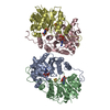
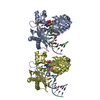
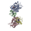
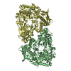
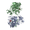
| Title | Model of gC1q-Fc complex based on 7A EM map | ||||||
 Components Components |
| ||||||
 Keywords Keywords | IMMUNE SYSTEM / Complement / antibody / complex / C1 | ||||||
| Function / homology |  Function and homology information Function and homology informationcomplement component C1q complex / IgM binding / complement component C1 complex / extrinsic component of postsynaptic membrane / negative regulation of macrophage differentiation / synapse pruning / extrinsic component of presynaptic membrane / negative regulation of granulocyte differentiation / vertebrate eye-specific patterning / complement-mediated synapse pruning ...complement component C1q complex / IgM binding / complement component C1 complex / extrinsic component of postsynaptic membrane / negative regulation of macrophage differentiation / synapse pruning / extrinsic component of presynaptic membrane / negative regulation of granulocyte differentiation / vertebrate eye-specific patterning / complement-mediated synapse pruning / symbiont cell surface / Dengue virus activates/modulates innate and adaptive immune responses / complement activation / IgG binding / Classical antibody-mediated complement activation / Initial triggering of complement / phosphatidylserine binding / neuron remodeling / complement activation, classical pathway / immunoglobulin complex / astrocyte activation / Regulation of Complement cascade / microglial cell activation / synapse organization / cell-cell signaling / amyloid-beta binding / extracellular matrix / blood microparticle / adaptive immune response / postsynapse / immune response / innate immune response / synapse / glutamatergic synapse / cell surface / : / extracellular region / plasma membrane Similarity search - Function | ||||||
| Biological species |  Homo sapiens (human) Homo sapiens (human) | ||||||
| Method | ELECTRON MICROSCOPY / single particle reconstruction / cryo EM / Resolution: 10 Å | ||||||
 Authors Authors | Ugurlar, D. / Howes, S.C. / de Kreuk, B.J.K. / de Jong, R.N. / Beurskens, F.J. / Koster, A.J. / Parren, P.W.H.I. / Sharp, T.H. / Gros, P. / Koning, R.I. | ||||||
| Funding support |  Netherlands, 1items Netherlands, 1items
| ||||||
 Citation Citation | Journal: Science / Year: 2018 Title: Structures of C1-IgG1 provide insights into how danger pattern recognition activates complement. Authors: Deniz Ugurlar / Stuart C Howes / Bart-Jan de Kreuk / Roman I Koning / Rob N de Jong / Frank J Beurskens / Janine Schuurman / Abraham J Koster / Thomas H Sharp / Paul W H I Parren / Piet Gros / Abstract: Danger patterns on microbes or damaged host cells bind and activate C1, inducing innate immune responses and clearance through the complement cascade. How these patterns trigger complement initiation ...Danger patterns on microbes or damaged host cells bind and activate C1, inducing innate immune responses and clearance through the complement cascade. How these patterns trigger complement initiation remains elusive. Here, we present cryo-electron microscopy analyses of C1 bound to monoclonal antibodies in which we observed heterogeneous structures of single and clustered C1-immunoglobulin G1 (IgG1) hexamer complexes. Distinct C1q binding sites are observed on the two Fc-CH2 domains of each IgG molecule. These are consistent with known interactions and also reveal additional interactions, which are supported by functional IgG1-mutant analysis. Upon antibody binding, the C1q arms condense, inducing rearrangements of the C1rs proteases and tilting C1q's cone-shaped stalk. The data suggest that C1r may activate C1s within single, strained C1 complexes or between neighboring C1 complexes on surfaces. | ||||||
| History |
|
- Structure visualization
| Movie |
Movie viewer |
|---|---|
| Structure viewer | Molecule: MolmilJmol/JSmol |
- Downloads & links
-Download
| PDBx/mmCIF format | 6fcz.cif.gz | 175 KB | Display | PDBx/mmCIF format |
|---|---|---|---|---|
| PDB format | pdb6fcz.ent.gz | 137.1 KB | Display | PDB format |
| PDBx/mmJSON format | 6fcz.json.gz | Tree view | PDBx/mmJSON format | |
| Others |  Other downloads Other downloads |
-Validation report
| Arichive directory | https://data.pdbj.org/pub/pdb/validation_reports/fc/6fczftp://data.pdbj.org/pub/pdb/validation_reports/fc/6fcz | HTTPS FTP |
|---|
-Related structure data
| Related structure data | 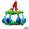 4232MC 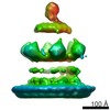 4231C C: citing same article ( M: map data used to model this data |
|---|---|
| Similar structure data |


-Links
 PDBj
PDBj


- Assembly
| Deposited unit | 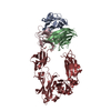
|
|---|---|
| 1 |
|
-Components
| #1: Protein | Mass: 14914.804 Da / Num. of mol.: 1 / Fragment: UNP residues 112-244 / Source method: isolated from a natural source / Source: (natural) Homo sapiens (human) / References: UniProt: P02745 | ||||
|---|---|---|---|---|---|
| #2: Protein | Mass: 14996.058 Da / Num. of mol.: 1 / Fragment: UNP Residues 119-250 / Source method: isolated from a natural source / Source: (natural) Homo sapiens (human) / References: UniProt: P02746 | ||||
| #3: Protein | Mass: 14302.143 Da / Num. of mol.: 1 / Fragment: UNP residues 117-245 / Source method: isolated from a natural source / Source: (natural) Homo sapiens (human) / References: UniProt: P02747 | ||||
| #4: Antibody | Mass: 24427.619 Da / Num. of mol.: 2 / Fragment: UNP residues 234-449 Source method: isolated from a genetically manipulated source Source: (gene. exp.) Homo sapiens (human) / Production host: Homo sapiens (human) / References: UniProt: P0DOX5#5: Water | ChemComp-HOH / |  Mass: 18.015 Da / Num. of mol.: 198 / Source method: isolated from a natural source / Formula: H2O Mass: 18.015 Da / Num. of mol.: 198 / Source method: isolated from a natural source / Formula: H2OHas protein modification | Y | |
-Experimental details
-Experiment
| Experiment | Method: ELECTRON MICROSCOPY |
|---|---|
| EM experiment | Aggregation state: PARTICLE / 3D reconstruction method: single particle reconstruction |
- Sample preparation
| Component |
| ||||||||||||||||||||||||
|---|---|---|---|---|---|---|---|---|---|---|---|---|---|---|---|---|---|---|---|---|---|---|---|---|---|
| Source (natural) |
| ||||||||||||||||||||||||
| Source (recombinant) | Organism: Homo sapiens (human) | ||||||||||||||||||||||||
| Buffer solution | pH: 7.5 | ||||||||||||||||||||||||
| Specimen | Embedding applied: NO / Shadowing applied: NO / Staining applied: NO / Vitrification applied: YES | ||||||||||||||||||||||||
| Vitrification | Cryogen name: ETHANE / Humidity: 100 % |
- Electron microscopy imaging
| Experimental equipment |  Model: Talos Arctica / Image courtesy: FEI Company |
|---|---|
| Microscopy | Model: FEI TALOS ARCTICA |
| Electron gun | Electron source:  FIELD EMISSION GUN / Accelerating voltage: 200 kV / Illumination mode: FLOOD BEAM FIELD EMISSION GUN / Accelerating voltage: 200 kV / Illumination mode: FLOOD BEAM |
| Electron lens | Mode: OTHER |
| Image recording | Electron dose: 40 e/Å2 / Film or detector model: FEI FALCON II (4k x 4k) |
- Processing
| CTF correction | Type: PHASE FLIPPING AND AMPLITUDE CORRECTION |
|---|---|
| Symmetry | Point symmetry: C1 (asymmetric) |
| 3D reconstruction | Resolution: 10 Å / Resolution method: FSC 0.143 CUT-OFF / Num. of particles: 79120 / Symmetry type: POINT |
| Atomic model building | Protocol: RIGID BODY FIT |
