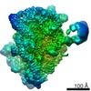
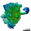
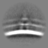
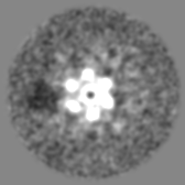
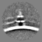
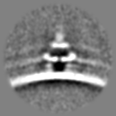
Journal: Science / Year: 2018 Title: Structures of C1-IgG1 provide insights into how danger pattern recognition activates complement. Authors: Deniz Ugurlar / Stuart C Howes / Bart-Jan de Kreuk / Roman I Koning / Rob N de Jong / Frank J Beurskens / Janine Schuurman / Abraham J Koster / Thomas H Sharp / Paul W H I Parren / Piet Gros / Abstract: Danger patterns on microbes or damaged host cells bind and activate C1, inducing innate immune responses and clearance through the complement cascade. How these patterns trigger complement initiation ...Danger patterns on microbes or damaged host cells bind and activate C1, inducing innate immune responses and clearance through the complement cascade. How these patterns trigger complement initiation remains elusive. Here, we present cryo-electron microscopy analyses of C1 bound to monoclonal antibodies in which we observed heterogeneous structures of single and clustered C1-immunoglobulin G1 (IgG1) hexamer complexes. Distinct C1q binding sites are observed on the two Fc-CH2 domains of each IgG molecule. These are consistent with known interactions and also reveal additional interactions, which are supported by functional IgG1-mutant analysis. Upon antibody binding, the C1q arms condense, inducing rearrangements of the C1rs proteases and tilting C1q's cone-shaped stalk. The data suggest that C1r may activate C1s within single, strained C1 complexes or between neighboring C1 complexes on surfaces.
History
Deposition
Dec 20, 2017
-
Header (metadata) release
Dec 27, 2017
-
Map release
Feb 28, 2018
-
Update
Sep 30, 2020
-
Current status
Sep 30, 2020
Processing site: PDBe / Status: Released
-
Structure visualization
Movie
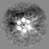
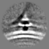
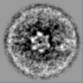
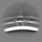
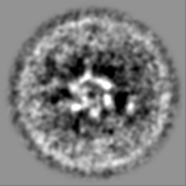
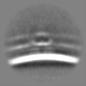
Surface view with section colored by density value
Cryogen name: ETHANE / Chamber humidity: 96 % / Chamber temperature: 294 K / Instrument: LEICA EM GP Details: 3 microlitres applied and incubated for 30 seconds, blot for 1 second before plunging.
Details
Extruded liposomes were incubated with IgG1 and C1. Gold fiducials were added just before applying to grids.
-
Electron microscopy
Microscope
FEI TITAN KRIOS
Specialist optics
Phase plate: VOLTA PHASE PLATE / Energy filter - Name: GIF Quantum LS
Image recording
Film or detector model: GATAN K2 SUMMIT (4k x 4k) / Detector mode: COUNTING / Digitization - Dimensions - Width: 3710 pixel / Digitization - Dimensions - Height: 3838 pixel / Digitization - Sampling interval: 5.0 µm / Digitization - Frames/image: 1-6 / Average electron dose: 1.2 e/Å2
Electron beam
Acceleration voltage: 300 kV / Electron source: FIELD EMISSION GUN
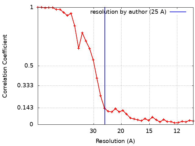
Applied symmetry - Point group: C1 (asymmetric) / Algorithm: BACK PROJECTION / Resolution.type: BY AUTHOR / Resolution: 25.0 Å / Resolution method: FSC 0.143 CUT-OFF / Software - Name: Dynamo / Number subtomograms used: 1660
Extraction
Number tomograms: 34 / Number images used: 51274 / Method: Manually annotate liposome surfaces / Software - Name: Dynamo (ver. 1.1.291) Details: Equally spaced sub-volumes were extracted from the surfaces of liposomes
Final 3D classification
Software - Name: Dynamo (ver. 1.1.291)
Final angle assignment
Type: OTHER / Software - Name: Dynamo (ver. 1.1.291) Details: Volume matching after filtering reference volume to angular range present in sub-volume
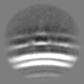
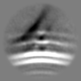
FSC plot (resolution estimation)
+
About Yorodumi
-
News
-
Feb 9, 2022. New format data for meta-information of EMDB entries
New format data for meta-information of EMDB entries
Version 3 of the EMDB header file is now the official format.
The previous official version 1.9 will be removed from the archive.
In the structure databanks used in Yorodumi, some data are registered as the other names, "COVID-19 virus" and "2019-nCoV". Here are the details of the virus and the list of structure data.
Jan 31, 2019. EMDB accession codes are about to change! (news from PDBe EMDB page)
EMDB accession codes are about to change! (news from PDBe EMDB page)
The allocation of 4 digits for EMDB accession codes will soon come to an end. Whilst these codes will remain in use, new EMDB accession codes will include an additional digit and will expand incrementally as the available range of codes is exhausted. The current 4-digit format prefixed with “EMD-” (i.e. EMD-XXXX) will advance to a 5-digit format (i.e. EMD-XXXXX), and so on. It is currently estimated that the 4-digit codes will be depleted around Spring 2019, at which point the 5-digit format will come into force.
The EM Navigator/Yorodumi systems omit the EMD- prefix.
Related info.:Q: What is EMD? / ID/Accession-code notation in Yorodumi/EM Navigator
Yorodumi is a browser for structure data from EMDB, PDB, SASBDB, etc.
This page is also the successor to EM Navigator detail page, and also detail information page/front-end page for Omokage search.
The word "yorodu" (or yorozu) is an old Japanese word meaning "ten thousand". "mi" (miru) is to see.
Related info.:EMDB / PDB / SASBDB / Comparison of 3 databanks / Yorodumi Search / Aug 31, 2016. New EM Navigator & Yorodumi / Yorodumi Papers / Jmol/JSmol / Function and homology information / Changes in new EM Navigator and Yorodumi
 Movie
Movie Controller


 Open data
 Basic information
 Map data
 Sample
 Function and homology information
 Homo sapiens (human)
 Authors
 Netherlands, 6 items
  Citation
 Structure visualization
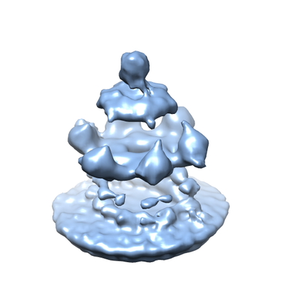
 Downloads & links
 emd_4231.png
 http://ftp.pdbj.org/pub/emdb/structures/EMD-4231


 Z (Sec.)
 Y (Row.)
 X (Col.)


 Sample components
 Processing
 Electron microscopy
 FIELD EMISSION GUN