| Entry | Database: PDB / ID: 6faj
|
|---|
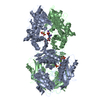
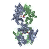
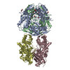
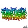
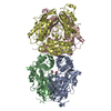
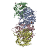
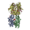
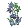
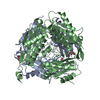
| Title | The structure of Human Methionine Adenosyltransferase II in apo state |
|---|
 Components Components | S-adenosylmethionine synthase isoform type-2 |
|---|
 Keywords Keywords | TRANSFERASE |
|---|
| Function / homology |  Function and homology information Function and homology information
methionine adenosyltransferase complex / methionine adenosyltransferase / methionine adenosyltransferase activity / S-adenosylmethionine biosynthetic process / protein heterooligomerization / Methylation / cellular response to methionine / protein hexamerization / small molecule binding / cellular response to leukemia inhibitory factor ...methionine adenosyltransferase complex / methionine adenosyltransferase / methionine adenosyltransferase activity / S-adenosylmethionine biosynthetic process / protein heterooligomerization / Methylation / cellular response to methionine / protein hexamerization / small molecule binding / cellular response to leukemia inhibitory factor / one-carbon metabolic process / positive regulation of TORC1 signaling / ATP binding / metal ion binding / identical protein binding / cytosolSimilarity search - Function GMP Synthetase; Chain A, domain 3 - #10 / S-adenosylmethionine synthetase / S-adenosylmethionine synthetase, N-terminal / S-adenosylmethionine synthetase, central domain / S-adenosylmethionine synthetase, C-terminal / S-adenosylmethionine synthetase, conserved site / S-adenosylmethionine synthetase superfamily / S-adenosylmethionine synthetase, N-terminal domain / S-adenosylmethionine synthetase, central domain / S-adenosylmethionine synthetase, C-terminal domain ...GMP Synthetase; Chain A, domain 3 - #10 / S-adenosylmethionine synthetase / S-adenosylmethionine synthetase, N-terminal / S-adenosylmethionine synthetase, central domain / S-adenosylmethionine synthetase, C-terminal / S-adenosylmethionine synthetase, conserved site / S-adenosylmethionine synthetase superfamily / S-adenosylmethionine synthetase, N-terminal domain / S-adenosylmethionine synthetase, central domain / S-adenosylmethionine synthetase, C-terminal domain / S-adenosylmethionine synthase signature 1. / S-adenosylmethionine synthase signature 2. / GMP Synthetase; Chain A, domain 3 / 2-Layer Sandwich / Alpha BetaSimilarity search - Domain/homology |
|---|
| Biological species |  Homo sapiens (human) Homo sapiens (human) |
|---|
| Method |  X-RAY DIFFRACTION / SYNCHROTRON / MOLECULAR REPLACEMENT / Resolution: 1.95 Å X-RAY DIFFRACTION / SYNCHROTRON / MOLECULAR REPLACEMENT / Resolution: 1.95 Å |
|---|
 Authors Authors | Bradley-Clarke, J. / Panmanee, J. / Antonyuk, S.V. / Hasnain, S.S. |
|---|
 Citation Citation | Journal: Febs J. / Year: 2020
Title: Control and regulation of S-Adenosylmethionine biosynthesis by the regulatory beta subunit and quinolone-based compounds
Authors: Panmanee, J. / Bradley-Clarke, J. / Mato, J.M. / O'Neill, P.M. / Antonyuk, S.V. / Hasnain, S.S. |
|---|
| History | | Deposition | Dec 15, 2017 | Deposition site: PDBE / Processing site: PDBE |
|---|
| Revision 1.0 | Feb 6, 2019 | Provider: repository / Type: Initial release |
|---|
| Revision 1.1 | Aug 19, 2020 | Group: Database references / Category: citation / citation_author
Item: _citation.country / _citation.journal_abbrev ..._citation.country / _citation.journal_abbrev / _citation.journal_id_CSD / _citation.journal_id_ISSN / _citation.pdbx_database_id_DOI / _citation.title / _citation.year |
|---|
| Revision 1.2 | Jan 17, 2024 | Group: Data collection / Database references / Refinement description
Category: chem_comp_atom / chem_comp_bond ...chem_comp_atom / chem_comp_bond / database_2 / pdbx_initial_refinement_model
Item: _database_2.pdbx_DOI / _database_2.pdbx_database_accession |
|---|
|
|---|
 Movie
Movie Controller

 Yorodumi
 Open data
 Basic information
 Structure visualization
 Downloads & links
 Other downloads
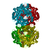


 PDBj
PDBj Assembly


 Mass: 106.120 Da / Num. of mol.: 2 / Source method: obtained synthetically / Formula: C4H10O3
 Mass: 194.226 Da / Num. of mol.: 1 / Source method: obtained synthetically / Formula: C8H18O5 / Comment: precipitant*YM
 Mass: 59.044 Da / Num. of mol.: 1 / Source method: obtained synthetically / Formula: C2H3O2
 Mass: 18.015 Da / Num. of mol.: 302 / Source method: isolated from a natural source / Formula: H2O
 Sample preparation
 / Beamline: I24 / Wavelength: 0.98 Å
 Processing