 Movie
Movie Controller

[English] 日本語
 Yorodumi
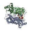
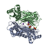
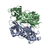
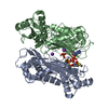
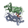
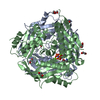
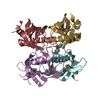
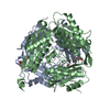
- PDB-6sw5: Crystal structure of the human S-adenosylmethionine synthetase 1 ... -
+ Open data

- Basic information
| Entry | Database: PDB / ID: 6sw5 | ||||||
|---|---|---|---|---|---|---|---|
| Title | Crystal structure of the human S-adenosylmethionine synthetase 1 (ligand-free form) | ||||||
 Components Components | S-adenosylmethionine synthase isoform type-1 | ||||||
 Keywords Keywords | TRANSFERASE / S-adenosylmethionine synthesis | ||||||
| Function / homology |  Function and homology information Function and homology informationDefective MAT1A causes MATD / L-methionine catabolic process / Sulfur amino acid metabolism / methionine adenosyltransferase complex / Metabolism of ingested SeMet, Sec, MeSec into H2Se / methionine adenosyltransferase / methionine adenosyltransferase activity / S-adenosylmethionine biosynthetic process / Methylation / one-carbon metabolic process ...Defective MAT1A causes MATD / L-methionine catabolic process / Sulfur amino acid metabolism / methionine adenosyltransferase complex / Metabolism of ingested SeMet, Sec, MeSec into H2Se / methionine adenosyltransferase / methionine adenosyltransferase activity / S-adenosylmethionine biosynthetic process / Methylation / one-carbon metabolic process / protein homotetramerization / ATP binding / metal ion binding / identical protein binding / cytosol Similarity search - Function | ||||||
| Biological species |  Homo sapiens (human) Homo sapiens (human) | ||||||
| Method |  X-RAY DIFFRACTION / SYNCHROTRON / MOLECULAR REPLACEMENT / Resolution: 2.35 Å X-RAY DIFFRACTION / SYNCHROTRON / MOLECULAR REPLACEMENT / Resolution: 2.35 Å | ||||||
 Authors Authors | Panmanee, J. / Antoyuk, S.V. / Hasnain, S.S. | ||||||
 Citation Citation | Journal: Acta Crystallogr D Struct Biol / Year: 2020 Title: Structural basis of the dominant inheritance of hypermethioninemia associated with the Arg264His mutation in the MAT1A gene. Authors: Panmanee, J. / Antonyuk, S.V. / Hasnain, S.S. | ||||||
| History |
|
- Structure visualization
| Structure viewer | Molecule: MolmilJmol/JSmol |
|---|
- Downloads & links
-Download
| PDBx/mmCIF format | 6sw5.cif.gz | 292.2 KB | Display | PDBx/mmCIF format |
|---|---|---|---|---|
| PDB format | pdb6sw5.ent.gz | 236.7 KB | Display | PDB format |
| PDBx/mmJSON format | 6sw5.json.gz | Tree view | PDBx/mmJSON format | |
| Others |  Other downloads Other downloads |
-Validation report
| Arichive directory | https://data.pdbj.org/pub/pdb/validation_reports/sw/6sw5ftp://data.pdbj.org/pub/pdb/validation_reports/sw/6sw5 | HTTPS FTP |
|---|
-Related structure data
| Related structure data | 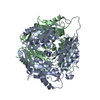 6sw6C 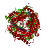 2obvS S: Starting model for refinement C: citing same article ( |
|---|---|
| Similar structure data |


-Links
 PDBj
PDBj- Assembly
| Deposited unit | 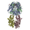
| ||||||||
|---|---|---|---|---|---|---|---|---|---|
| 1 | 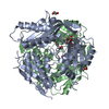
| ||||||||
| 2 | 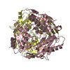
| ||||||||
| Unit cell |
|
-Components
| #1: Protein | Mass: 43703.852 Da / Num. of mol.: 4 Source method: isolated from a genetically manipulated source Source: (gene. exp.) Homo sapiens (human) / Tissue: Liver / Cell: Hepatocyte / Gene: MAT1A, AMS1, MATA1 / Organ: Liver / Plasmid: pNIC28-Bsa4 / Production host:  #2: Chemical |   Mass: 106.120 Da / Num. of mol.: 2 / Source method: obtained synthetically / Formula: C4H10O3 Mass: 106.120 Da / Num. of mol.: 2 / Source method: obtained synthetically / Formula: C4H10O3#3: Chemical | ChemComp-EDO /   Mass: 62.068 Da / Num. of mol.: 12 / Source method: obtained synthetically / Formula: C2H6O2 Mass: 62.068 Da / Num. of mol.: 12 / Source method: obtained synthetically / Formula: C2H6O2#4: Water | ChemComp-HOH / |  Mass: 18.015 Da / Num. of mol.: 399 / Source method: isolated from a natural source / Formula: H2O Mass: 18.015 Da / Num. of mol.: 399 / Source method: isolated from a natural source / Formula: H2OHas ligand of interest | N | |
|---|
-Experimental details
-Experiment
| Experiment | Method: X-RAY DIFFRACTION / Number of used crystals: 1 |
|---|
- Sample preparation
| Crystal | Density Matthews: 2.49 Å3/Da / Density % sol: 50.56 % / Description: Plate shape |
|---|---|
| Crystal grow | Temperature: 298 K / Method: vapor diffusion, hanging drop / pH: 7.5 / Details: 200 mM NaF, 20 % PEG 3350 and 15 % ethylene glycol |
-Data collection
| Diffraction | Mean temperature: 100 K / Serial crystal experiment: N |
|---|---|
| Diffraction source | Source: SYNCHROTRON / Site: Diamond  / Beamline: I04 / Wavelength: 0.9795 Å / Beamline: I04 / Wavelength: 0.9795 Å |
| Detector | Type: DECTRIS EIGER2 X 16M / Detector: PIXEL / Date: Jun 29, 2019 |
| Radiation | Protocol: SINGLE WAVELENGTH / Monochromatic (M) / Laue (L): M / Scattering type: x-ray |
| Radiation wavelength | Wavelength: 0.9795 Å / Relative weight: 1 |
| Reflection | Resolution: 2.35→58.76 Å / Num. obs: 66266 / % possible obs: 100 % / Redundancy: 3.4 % / Biso Wilson estimate: 29.335 Å2 / CC1/2: 0.991 / Rmerge(I) obs: 0.127 / Rpim(I) all: 0.08 / Rrim(I) all: 0.15 / Net I/σ(I): 6.4 |
| Reflection shell | Resolution: 2.35→2.41 Å / Redundancy: 3.4 % / Rmerge(I) obs: 0.58 / Mean I/σ(I) obs: 1.9 / Num. unique obs: 4425 / CC1/2: 0.762 / Rpim(I) all: 0.365 / Rrim(I) all: 0.686 / % possible all: 99.9 |
- Processing
| Software |
| ||||||||||||||||||||||||||||||||||||||||||||||||||||||||||||
|---|---|---|---|---|---|---|---|---|---|---|---|---|---|---|---|---|---|---|---|---|---|---|---|---|---|---|---|---|---|---|---|---|---|---|---|---|---|---|---|---|---|---|---|---|---|---|---|---|---|---|---|---|---|---|---|---|---|---|---|---|---|
| Refinement | Method to determine structure: MOLECULAR REPLACEMENT Starting model: 2OBV Resolution: 2.35→58.76 Å / Cor.coef. Fo:Fc: 0.949 / Cor.coef. Fo:Fc free: 0.92 / SU B: 8.642 / SU ML: 0.195 / Cross valid method: THROUGHOUT / σ(F): 0 / ESU R: 0.383 / ESU R Free: 0.246 Details: HYDROGENS HAVE BEEN ADDED IN THE RIDING POSITIONS U VALUES : REFINED INDIVIDUALLY
| ||||||||||||||||||||||||||||||||||||||||||||||||||||||||||||
| Solvent computation | Ion probe radii: 0.8 Å / Shrinkage radii: 0.8 Å / VDW probe radii: 1.2 Å | ||||||||||||||||||||||||||||||||||||||||||||||||||||||||||||
| Displacement parameters | Biso max: 127.88 Å2 / Biso mean: 35.909 Å2 / Biso min: 1 Å2
| ||||||||||||||||||||||||||||||||||||||||||||||||||||||||||||
| Refinement step | Cycle: final / Resolution: 2.35→58.76 Å
| ||||||||||||||||||||||||||||||||||||||||||||||||||||||||||||
| Refine LS restraints |
| ||||||||||||||||||||||||||||||||||||||||||||||||||||||||||||
| LS refinement shell | Resolution: 2.35→2.411 Å / Rfactor Rfree error: 0 / Total num. of bins used: 20
|
