 Movie
Movie Controller

[English] 日本語
 Yorodumi
- PDB-1qm4: Methionine Adenosyltransferase Complexed with a L-Methionine Analogue -
+ Open data

- Basic information
| Entry | Database: PDB / ID: 1qm4 | ||||||
|---|---|---|---|---|---|---|---|
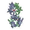
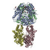
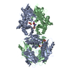
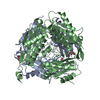
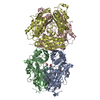
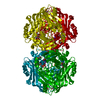
| Title | Methionine Adenosyltransferase Complexed with a L-Methionine Analogue | ||||||
 Components Components | METHIONINE ADENOSYLTRANSFERASE, ALPHA FORM | ||||||
 Keywords Keywords | TRANSFERASE / ADENOSYLTRANSFERASE / METHIONINE BINDING | ||||||
| Function / homology |  Function and homology information Function and homology informationMetabolism of ingested SeMet, Sec, MeSec into H2Se / Sulfur amino acid metabolism / L-methionine catabolic process / methionine adenosyltransferase complex / Methylation / methionine adenosyltransferase / methionine adenosyltransferase activity / S-adenosylmethionine biosynthetic process / amino acid binding / one-carbon metabolic process ...Metabolism of ingested SeMet, Sec, MeSec into H2Se / Sulfur amino acid metabolism / L-methionine catabolic process / methionine adenosyltransferase complex / Methylation / methionine adenosyltransferase / methionine adenosyltransferase activity / S-adenosylmethionine biosynthetic process / amino acid binding / one-carbon metabolic process / ADP binding / nuclear matrix / protein-containing complex assembly / protein homotetramerization / magnesium ion binding / ATP binding / identical protein binding / cytosol Similarity search - Function | ||||||
| Biological species |  | ||||||
| Method |  X-RAY DIFFRACTION / MOLECULAR REPLACEMENT / Resolution: 2.66 Å X-RAY DIFFRACTION / MOLECULAR REPLACEMENT / Resolution: 2.66 Å | ||||||
 Authors Authors | Gonzalez, B. / Pajares, M.A. / Hermoso, J.A. / Sanz-Aparicio, J. | ||||||
 Citation Citation | Journal: J.Mol.Biol. / Year: 2000 Title: The Crystal Structure of Tetrameric Methionine Adenosyltransferase from Rat Liver Reveals the Methionine-Binding Site Authors: Gonzalez, B. / Pajares, M.A. / Hermoso, J.A. / Alvarez, L. / Garrido, F. / Sufrin, J.R. / Sanz-Aparicio, J. #1: Journal: Biochem.J. / Year: 1994 Title: Expression of Rat Liver S-Adenosylmethionine Synthetase in Escherichia Coli Results in Two Active Oligomeric Forms Authors: Alvarez, L. / Mingorance, J. / Pajares, M.A. / Mato, J.M. | ||||||
| History |
|
- Structure visualization
| Structure viewer | Molecule: MolmilJmol/JSmol |
|---|
- Downloads & links
-Download
| PDBx/mmCIF format | 1qm4.cif.gz | 157.5 KB | Display | PDBx/mmCIF format |
|---|---|---|---|---|
| PDB format | pdb1qm4.ent.gz | 123.2 KB | Display | PDB format |
| PDBx/mmJSON format | 1qm4.json.gz | Tree view | PDBx/mmJSON format | |
| Others |  Other downloads Other downloads |
-Validation report
| Arichive directory | https://data.pdbj.org/pub/pdb/validation_reports/qm/1qm4ftp://data.pdbj.org/pub/pdb/validation_reports/qm/1qm4 | HTTPS FTP |
|---|
-Related structure data
| Related structure data | 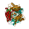 1mxaS S: Starting model for refinement |
|---|---|
| Similar structure data |


-Links
 PDBj
PDBj- Assembly
| Deposited unit | 
| ||||||||
|---|---|---|---|---|---|---|---|---|---|
| 1 | 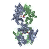
| ||||||||
| Unit cell |
| ||||||||
| Noncrystallographic symmetry (NCS) | NCS oper: (Code: given Matrix: (-0.78343, -0.23114, 0.5769), Vector: Details | THE ENZYME IS A TETRAMERTHE DIMER IN THE ASYMMETRIC UNIT HAS A PROTEIN PROTEININTERACTION ABOUT THE LARGEST FACE OF THE MONOMERICMOLECULE AND THE TETRAMER IS MADE UP OF TWO OF THESEDIMERS INTERACTING IN A 'HEAD-TO-HEAD' MANNER .IN THE CRYSTAL THE DIMER MOLECULES ARE INVOLVED IN A STRONGCRYSTAL PACKING INTERACTION INVOLVING THE OPPOSITE FACEOF THE MOLECULES TO THAT INVOLVED IN THE BIOLOGICAL DIMER.FOR EXAMPLE SYMMETRY OPERATION,BIOMT1 2 1.000000 0.000000 0. 000000 0.00000BIOMT2 2 0.000000 -1.000000 0.000000 115.20000BIOMT3 2 0.000000 0. 000000 -1.000000 79.99000GENERATES A CRYSTAL PACKING TETRAMERIC ASSEMBLY. | |
-Components
-Protein , 1 types, 2 molecules AB
| #1: Protein | Mass: 43637.734 Da / Num. of mol.: 2 Source method: isolated from a genetically manipulated source Source: (gene. exp.)  |
|---|
-Non-polymers , 5 types, 206 molecules 


| #2: Chemical | ChemComp-AMB /  Mass: 131.130 Da / Num. of mol.: 1 / Source method: obtained synthetically / Formula: C5H9NO3 Mass: 131.130 Da / Num. of mol.: 1 / Source method: obtained synthetically / Formula: C5H9NO3 | ||||||
|---|---|---|---|---|---|---|---|
| #3: Chemical |  Mass: 96.063 Da / Num. of mol.: 3 / Source method: obtained synthetically / Formula: SO4 Mass: 96.063 Da / Num. of mol.: 3 / Source method: obtained synthetically / Formula: SO4#4: Chemical |  Mass: 24.305 Da / Num. of mol.: 2 / Source method: obtained synthetically / Formula: Mg Mass: 24.305 Da / Num. of mol.: 2 / Source method: obtained synthetically / Formula: Mg#5: Chemical |  Mass: 39.098 Da / Num. of mol.: 3 / Source method: obtained synthetically / Formula: K Mass: 39.098 Da / Num. of mol.: 3 / Source method: obtained synthetically / Formula: K#6: Water | ChemComp-HOH / | Mass: 18.015 Da / Num. of mol.: 197 / Source method: isolated from a natural source / Formula: H2O |
-Experimental details
-Experiment
| Experiment | Method: X-RAY DIFFRACTION / Number of used crystals: 1 |
|---|
- Sample preparation
| Crystal | Density Matthews: 3.2 Å3/Da / Density % sol: 61 % | ||||||||||||||||||||||||||||||||||||||||||||||||||||||||||||||||||||||
|---|---|---|---|---|---|---|---|---|---|---|---|---|---|---|---|---|---|---|---|---|---|---|---|---|---|---|---|---|---|---|---|---|---|---|---|---|---|---|---|---|---|---|---|---|---|---|---|---|---|---|---|---|---|---|---|---|---|---|---|---|---|---|---|---|---|---|---|---|---|---|---|
| Crystal grow | pH: 7.5 Details: 16 % PEG 10K, 10MM CIS-AMB, 0.1 HEPES PH=7.5, pH 7.50 | ||||||||||||||||||||||||||||||||||||||||||||||||||||||||||||||||||||||
| Crystal grow | *PLUS pH: 8 / Method: vapor diffusion, hanging dropDetails: drop consists of equal amounts of protein and precipitant solutions | ||||||||||||||||||||||||||||||||||||||||||||||||||||||||||||||||||||||
| Components of the solutions | *PLUS
|
-Data collection
| Diffraction | Mean temperature: 120 K |
|---|---|
| Diffraction source | Source: ROTATING ANODE / Type: ENRAF-NONIUS / Wavelength: 1.5418 |
| Detector | Type: MARRESEARCH / Detector: IMAGE PLATE / Date: Feb 15, 1999 |
| Radiation | Monochromator: GRAPHITE(002) / Protocol: SINGLE WAVELENGTH / Monochromatic (M) / Laue (L): M / Scattering type: x-ray |
| Radiation wavelength | Wavelength: 1.5418 Å / Relative weight: 1 |
| Reflection | Resolution: 2.66→44.7 Å / Num. obs: 28742 / % possible obs: 94.5 % / Observed criterion σ(I): 4 / Redundancy: 7.5 % / Rmerge(I) obs: 0.082 / Rsym value: 0.082 / Net I/σ(I): 6.7 |
| Reflection shell | Resolution: 2.66→2.8 Å / Redundancy: 6.9 % / Mean I/σ(I) obs: 2.1 / Rsym value: 0.37 / % possible all: 85.8 |
| Reflection shell | *PLUS % possible obs: 85.8 % / Rmerge(I) obs: 0.37 |
- Processing
| Software |
| ||||||||||||||||||||||||||||||||||||||||||||||||||||||||||||
|---|---|---|---|---|---|---|---|---|---|---|---|---|---|---|---|---|---|---|---|---|---|---|---|---|---|---|---|---|---|---|---|---|---|---|---|---|---|---|---|---|---|---|---|---|---|---|---|---|---|---|---|---|---|---|---|---|---|---|---|---|---|
| Refinement | Method to determine structure: MOLECULAR REPLACEMENT Starting model: 1MXA Resolution: 2.66→10 Å / Data cutoff high absF: 10000000 / Data cutoff low absF: 0.001 / Isotropic thermal model: RESTRAINED / Cross valid method: THROUGHOUT / σ(F): 2 Details: NO DENSITY FOR RESIDUES A1-A16, AND B1-B16, WAS OBSERVED IN THE DENSITY MAPS
| ||||||||||||||||||||||||||||||||||||||||||||||||||||||||||||
| Displacement parameters | Biso mean: 41.5 Å2 | ||||||||||||||||||||||||||||||||||||||||||||||||||||||||||||
| Refinement step | Cycle: LAST / Resolution: 2.66→10 Å
| ||||||||||||||||||||||||||||||||||||||||||||||||||||||||||||
| Refine LS restraints |
| ||||||||||||||||||||||||||||||||||||||||||||||||||||||||||||
| LS refinement shell | Resolution: 2.66→2.78 Å / Total num. of bins used: 8
| ||||||||||||||||||||||||||||||||||||||||||||||||||||||||||||
| Xplor file |
| ||||||||||||||||||||||||||||||||||||||||||||||||||||||||||||
| Software | *PLUS Name: X-PLOR / Version: 3.843 / Classification: refinement | ||||||||||||||||||||||||||||||||||||||||||||||||||||||||||||
| Refine LS restraints | *PLUS
|
