[English] 日本語
Yorodumi
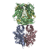
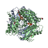
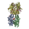
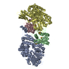
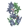
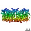
- PDB-3so4: Methionine-adenosyltransferase from Entamoeba histolytica -

+
Open data


ID or keywords:

Loading...

-
Basic information

Entry
Database: PDB / ID: 3so4
TitleMethionine-adenosyltransferase from Entamoeba histolytica
ComponentsMethionine-adenosyltransferase
KeywordsTRANSFERASE / Structural Genomics / Medical Structural Genomics of Pathogenic Protozoa / MSGPP / S-adenosylmethionine synthetase / PSI / Protein Structure Initiative
Function / homology
Function and homology information


methionine adenosyltransferase / methionine adenosyltransferase activity / S-adenosylmethionine biosynthetic process / one-carbon metabolic process / ATP binding / metal ion binding / cytosol
Similarity search - Function
GMP Synthetase; Chain A, domain 3 - #10 / S-adenosylmethionine synthetase / S-adenosylmethionine synthetase, N-terminal / S-adenosylmethionine synthetase, central domain / S-adenosylmethionine synthetase, C-terminal / S-adenosylmethionine synthetase, conserved site / S-adenosylmethionine synthetase superfamily / S-adenosylmethionine synthetase, N-terminal domain / S-adenosylmethionine synthetase, central domain / S-adenosylmethionine synthetase, C-terminal domain ...GMP Synthetase; Chain A, domain 3 - #10 / S-adenosylmethionine synthetase / S-adenosylmethionine synthetase, N-terminal / S-adenosylmethionine synthetase, central domain / S-adenosylmethionine synthetase, C-terminal / S-adenosylmethionine synthetase, conserved site / S-adenosylmethionine synthetase superfamily / S-adenosylmethionine synthetase, N-terminal domain / S-adenosylmethionine synthetase, central domain / S-adenosylmethionine synthetase, C-terminal domain / S-adenosylmethionine synthase signature 1. / S-adenosylmethionine synthase signature 2. / GMP Synthetase; Chain A, domain 3 / 2-Layer Sandwich / Alpha Beta
Similarity search - Domain/homology
ACETATE ION / S-adenosylmethionine synthase
Similarity search - Component
Biological speciesEntamoeba histolytica (eukaryote)
MethodX-RAY DIFFRACTION / SYNCHROTRON / MOLECULAR REPLACEMENT / molecular replacement / Resolution: 3.18 Å
AuthorsMerritt, E.A. / Medical Structural Genomics of Pathogenic Protozoa (MSGPP)
CitationJournal: To be Published
Title: Methionine-adenosyltransferase from Entamoeba histolytica
Authors: Merritt, E.A. / Arakaki, T. / Zhang, L. / Napuli, A. / Van Voorhis, W.C. / Buckner, F.S. / Fan, E. / Zucker, F. / Verlinde, C.L.M.J. / Hol, W.G.J.
History
DepositionJun 29, 2011Deposition site: RCSB / Processing site: RCSB
Revision 1.0Jul 20, 2011Provider: repository / Type: Initial release
Revision 1.1Nov 8, 2017Group: Refinement description / Category: software
Item: _software.classification / _software.contact_author ..._software.classification / _software.contact_author / _software.contact_author_email / _software.date / _software.language / _software.location / _software.name / _software.type / _software.version
Revision 1.2Feb 28, 2024Group: Data collection / Database references / Derived calculations
Category: chem_comp_atom / chem_comp_bond ...chem_comp_atom / chem_comp_bond / database_2 / struct_ref_seq_dif / struct_site
Item: _database_2.pdbx_DOI / _database_2.pdbx_database_accession ..._database_2.pdbx_DOI / _database_2.pdbx_database_accession / _struct_ref_seq_dif.details / _struct_site.pdbx_auth_asym_id / _struct_site.pdbx_auth_comp_id / _struct_site.pdbx_auth_seq_id

-
Structure visualization

Structure viewerMolecule:
MolmilJmol/JSmol

Downloads & links

-
Assembly

Deposited unit
A: Methionine-adenosyltransferase
B: Methionine-adenosyltransferase
C: Methionine-adenosyltransferase
D: Methionine-adenosyltransferase
hetero molecules


Theoretical massNumber of molelcules
Total (without water)182,9448
Polymers182,7084
Non-polymers2364
Water724
1


  • Idetical with deposited unit
  • defined by author&software
TypeNameSymmetry operationNumber
identity operation1_555x,y,z1
Buried area13930 Å2
ΔGint-58 kcal/mol
Surface area48720 Å2
MethodPISA
Unit cell
Length a, b, c (Å)58.492, 113.161, 220.848
Angle α, β, γ (deg.)90.000, 90.000, 90.000
Int Tables number19
Space group name H-MP212121
DetailsTetramer

-
Components

#1: Protein
Methionine-adenosyltransferase / S-adenosylmethionine synthase


Mass: 45677.023 Da / Num. of mol.: 4 / Fragment: residues 4-397
Source method: isolated from a genetically manipulated source
Source: (gene. exp.) Entamoeba histolytica (eukaryote) / Gene: 103.m00148, EHI_174250 / Plasmid: AVA0421 / Production host: Escherichia coli (E. coli) / References: UniProt: C4M272, methionine adenosyltransferase
#2: Chemical
ChemComp-ACT / ACETATE ION


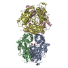
Mass: 59.044 Da / Num. of mol.: 4 / Source method: obtained synthetically / Formula: C2H3O2
#3: Water ChemComp-HOH / water


Mass: 18.015 Da / Num. of mol.: 4 / Source method: isolated from a natural source / Formula: H2O

-
Experimental details

-
Experiment

ExperimentMethod: X-RAY DIFFRACTION / Number of used crystals: 1

-
Sample preparation

CrystalDensity Matthews: 2 Å3/Da / Density % sol: 38.5 %
Crystal growTemperature: 293 K / Method: vapor diffusion / pH: 6.2
Details: protein buffer 25 mM HEPES pH 7.5, 500 mM NaCl; crystallization buffer 400 mM Ammonium Acetate, 100 mM BisTris pH 6.2, 33% PEG 3350, vapor diffusion, temperature 293K

-
Data collection

DiffractionMean temperature: 100 K
Diffraction sourceSource: SYNCHROTRON / Site: SSRL / Beamline: BL11-1 / Wavelength: 0.97945 Å
DetectorDetector: CCD / Date: May 21, 2008
RadiationProtocol: SINGLE WAVELENGTH / Monochromatic (M) / Laue (L): M / Scattering type: x-ray
Radiation wavelengthWavelength: 0.97945 Å / Relative weight: 1
ReflectionResolution: 3.18→50 Å / Num. obs: 23472 / % possible obs: 92.2 % / Redundancy: 3.8 % / Rmerge(I) obs: 0.171 / Χ2: 1.106 / Net I/σ(I): 4.8
Reflection shell
Resolution (Å)Redundancy (%)Rmerge(I) obsNum. unique allΧ2Diffraction-ID% possible all
3.2-3.272.50.7169420.999156
3.27-3.362.80.76211171.009168.5
3.36-3.452.90.59212921.004177.2
3.45-3.553.10.57314801.088189
3.55-3.663.40.48816101.043195.3
3.66-3.793.70.3616241.097198.1
3.79-3.953.90.37716751.077199.8
3.95-4.134.10.30116821.1211100
4.13-4.344.10.24216621.0951100
4.34-4.614.10.16917051.151100
4.61-4.974.20.16417121.1381100
4.97-5.474.20.14416921.114199.9
5.47-6.264.20.15517161.111199.9
6.26-7.884.20.09917501.115199.7
7.88-5040.0418131.196197

-
Phasing

PhasingMethod: molecular replacement

-
Processing

Software
NameVersionClassificationNB
DENZOdata reduction
SCALEPACKdata scaling
REFMACrefinement
PDB_EXTRACT3.1data extraction
Blu-Icedata collection
HKL-2000data reduction
BALBESphasing
RefinementMethod to determine structure: MOLECULAR REPLACEMENT / Resolution: 3.18→50 Å / Cor.coef. Fo:Fc: 0.919 / Cor.coef. Fo:Fc free: 0.871 / WRfactor Rfree: 0.2386 / WRfactor Rwork: 0.1919 / Occupancy max: 1 / Occupancy min: 1 / FOM work R set: 0.7982 / SU B: 75.438 / SU ML: 0.562 / SU R Cruickshank DPI: 0.5531 / SU Rfree: 0.6828 / Cross valid method: THROUGHOUT / σ(F): 0 / ESU R Free: 0.683 / Stereochemistry target values: MAXIMUM LIKELIHOOD
Details: HYDROGENS HAVE BEEN USED IF PRESENT IN THE INPUT U VALUES : WITH TLS ADDED
RfactorNum. reflection% reflectionSelection details
Rfree0.2839 1189 5.1 %RANDOM
Rwork0.2278 ---
obs0.2306 23302 91.13 %-
Solvent computationIon probe radii: 0.8 Å / Shrinkage radii: 0.8 Å / VDW probe radii: 1.2 Å / Solvent model: MASK
Displacement parametersBiso max: 147.96 Å2 / Biso mean: 80.1164 Å2 / Biso min: 24.63 Å2
Baniso -1Baniso -2Baniso -3
1-1.41 Å20 Å20 Å2
2--2.19 Å20 Å2
3----3.61 Å2
Refinement stepCycle: LAST / Resolution: 3.18→50 Å
ProteinNucleic acidLigandSolventTotal
Num. atoms11478 0 16 4 11498
Refine LS restraints
Refine-IDTypeDev idealDev ideal targetNumber
X-RAY DIFFRACTIONr_bond_refined_d0.0090.02211714
X-RAY DIFFRACTIONr_bond_other_d0.0050.027811
X-RAY DIFFRACTIONr_angle_refined_deg1.2031.94315802
X-RAY DIFFRACTIONr_angle_other_deg1.349319159
X-RAY DIFFRACTIONr_dihedral_angle_1_deg5.52251508
X-RAY DIFFRACTIONr_chiral_restr0.0660.21803
X-RAY DIFFRACTIONr_gen_planes_refined0.0040.0212836
X-RAY DIFFRACTIONr_gen_planes_other0.0030.022232
LS refinement shellResolution: 3.178→3.26 Å / Total num. of bins used: 20
RfactorNum. reflection% reflection
Rfree0.415 43 -
Rwork0.386 830 -
all-873 -
obs--48.69 %
Refinement TLS params.

Method: refined / Refine-ID: X-RAY DIFFRACTION

IDL112)L122)L132)L222)L232)L332)S11 (Å °)S12 (Å °)S13 (Å °)S21 (Å °)S22 (Å °)S23 (Å °)S31 (Å °)S32 (Å °)S33 (Å °)T112)T122)T132)T222)T232)T332)Origin x (Å)Origin y (Å)Origin z (Å)
11.2946-0.0905-0.08361.83130.15790.80550.09260.04280.09830.1923-0.1031-0.27050.01390.05410.01050.2010.0002-0.03270.22780.07240.078825.614-5.751-12.21
20.88130.23420.28091.86510.09041.3115-0.00260.37910.2265-0.0891-0.02740.0881-0.09960.03720.030.18630.0570.05490.34210.1310.08069.2191.405-28.581
30.5568-0.0023-0.45131.99580.24391.682-0.24330.1325-0.29410.0589-0.13710.18490.3876-0.09410.38040.3467-0.01230.18530.2174-0.1310.20931.916-55.03-28.381
41.25050.4090.04922.38550.80371.5326-0.17680.3272-0.171-0.42840.0138-0.3890.1440.1150.1630.40970.05140.20180.4435-0.06680.168720.244-52.606-44.323
Refinement TLS group
IDRefine-IDRefine TLS-IDAuth asym-IDAuth seq-ID
1X-RAY DIFFRACTION1A3 - 397
2X-RAY DIFFRACTION2B3 - 397
3X-RAY DIFFRACTION3C3 - 397
4X-RAY DIFFRACTION4D3 - 397

+
About Yorodumi

-
News

-
Feb 9, 2022. New format data for meta-information of EMDB entries

New format data for meta-information of EMDB entries

  • Version 3 of the EMDB header file is now the official format.
  • The previous official version 1.9 will be removed from the archive.

Related info.:EMDB header

External links:wwPDB to switch to version 3 of the EMDB data model

-
Aug 12, 2020. Covid-19 info

Covid-19 info

URL: https://pdbjlvh1.pdbj.org/emnavi/covid19.php

New page: Covid-19 featured information page in EM Navigator.

Related info.:Covid-19 info / Mar 5, 2020. Novel coronavirus structure data

+
Mar 5, 2020. Novel coronavirus structure data

Novel coronavirus structure data

Related info.:Yorodumi Speices / Aug 12, 2020. Covid-19 info

External links:COVID-19 featured content - PDBj / Molecule of the Month (242):Coronavirus Proteases

+
Jan 31, 2019. EMDB accession codes are about to change! (news from PDBe EMDB page)

EMDB accession codes are about to change! (news from PDBe EMDB page)

  • The allocation of 4 digits for EMDB accession codes will soon come to an end. Whilst these codes will remain in use, new EMDB accession codes will include an additional digit and will expand incrementally as the available range of codes is exhausted. The current 4-digit format prefixed with “EMD-” (i.e. EMD-XXXX) will advance to a 5-digit format (i.e. EMD-XXXXX), and so on. It is currently estimated that the 4-digit codes will be depleted around Spring 2019, at which point the 5-digit format will come into force.
  • The EM Navigator/Yorodumi systems omit the EMD- prefix.

Related info.:Q: What is EMD? / ID/Accession-code notation in Yorodumi/EM Navigator

External links:EMDB Accession Codes are Changing Soon! / Contact to PDBj

+
Jul 12, 2017. Major update of PDB

Major update of PDB

  • wwPDB released updated PDB data conforming to the new PDBx/mmCIF dictionary.
  • This is a major update changing the version number from 4 to 5, and with Remediation, in which all the entries are updated.
  • In this update, many items about electron microscopy experimental information are reorganized (e.g. em_software).
  • Now, EM Navigator and Yorodumi are based on the updated data.

External links:wwPDB Remediation / Enriched Model Files Conforming to OneDep Data Standards Now Available in the PDB FTP Archive

-
Yorodumi

Thousand views of thousand structures

  • Yorodumi is a browser for structure data from EMDB, PDB, SASBDB, etc.
  • This page is also the successor to EM Navigator detail page, and also detail information page/front-end page for Omokage search.
  • The word "yorodu" (or yorozu) is an old Japanese word meaning "ten thousand". "mi" (miru) is to see.

Related info.:EMDB / PDB / SASBDB / Comparison of 3 databanks / Yorodumi Search / Aug 31, 2016. New EM Navigator & Yorodumi / Yorodumi Papers / Jmol/JSmol / Function and homology information / Changes in new EM Navigator and Yorodumi

Read more