 Movie
Movie Controller

[English] 日本語
 Yorodumi
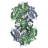
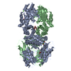
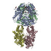
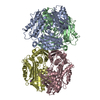
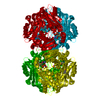
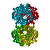
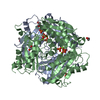
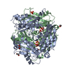
- PDB-1o9t: Methionine adenosyltransferase complexed with both substrates ATP... -
+ Open data

- Basic information
| Entry | Database: PDB / ID: 1o9t | ||||||
|---|---|---|---|---|---|---|---|
| Title | Methionine adenosyltransferase complexed with both substrates ATP and methionine | ||||||
 Components Components | S-ADENOSYLMETHIONINE SYNTHETASE | ||||||
 Keywords Keywords | TRANSFERASE / ADENOSYLTRANSFERASE / ATP BINDING / METHIONINE BINDING / ONE-CARBON METABOLISM / MAGNESIUM / POTASSIUM / METAL-BINDING / MULTIGENE FAMILY / ATP-BINDING | ||||||
| Function / homology |  Function and homology information Function and homology informationMetabolism of ingested SeMet, Sec, MeSec into H2Se / Sulfur amino acid metabolism / L-methionine catabolic process / methionine adenosyltransferase complex / Methylation / methionine adenosyltransferase / methionine adenosyltransferase activity / S-adenosylmethionine biosynthetic process / amino acid binding / one-carbon metabolic process ...Metabolism of ingested SeMet, Sec, MeSec into H2Se / Sulfur amino acid metabolism / L-methionine catabolic process / methionine adenosyltransferase complex / Methylation / methionine adenosyltransferase / methionine adenosyltransferase activity / S-adenosylmethionine biosynthetic process / amino acid binding / one-carbon metabolic process / ADP binding / nuclear matrix / protein-containing complex assembly / protein homotetramerization / magnesium ion binding / ATP binding / identical protein binding / cytosol Similarity search - Function | ||||||
| Biological species |  | ||||||
| Method |  X-RAY DIFFRACTION / MOLECULAR REPLACEMENT / Resolution: 2.9 Å X-RAY DIFFRACTION / MOLECULAR REPLACEMENT / Resolution: 2.9 Å | ||||||
 Authors Authors | Gonzalez, B. / Pajares, M.A. / Hermoso, J.A. / Sanz-Aparicio, J. | ||||||
 Citation Citation | Journal: J.Mol.Biol. / Year: 2003 Title: Crystal Structures of Methionine Adenosyltransferase Complexed with Substrates and Products Reveal the Methionine-ATP Recognition and Give Insights Into the Catalytic Mechanism Authors: Gonzalez, B. / Pajares, M.A. / Hermoso, J.A. / Guillerm, D. / Guillerm, G. / Sanz-Aparicio, J. #1: Journal: J.Mol.Biol. / Year: 2000Title: The Crystal Structure of Tetrameric Methionine Adenosyltransferase Fom Rat Liver Reveals the Methionine Binding Site Authors: Gonzalez, B. / Pajares, M.A. / Hermoso, J.A. / Alvarez, L. / Garrido, F. / Sufrin, J.R. / Sanz-Aparicio, J. | ||||||
| History |
| ||||||
| Remark 700 | SHEET THE SHEET STRUCTURE OF THIS MOLECULE IS BIFURCATED. IN ORDER TO REPRESENT THIS FEATURE IN ... SHEET THE SHEET STRUCTURE OF THIS MOLECULE IS BIFURCATED. IN ORDER TO REPRESENT THIS FEATURE IN THE SHEET RECORDS BELOW, TWO SHEETS ARE DEFINED. |
- Structure visualization
| Structure viewer | Molecule: MolmilJmol/JSmol |
|---|
- Downloads & links
-Download
| PDBx/mmCIF format | 1o9t.cif.gz | 159.3 KB | Display | PDBx/mmCIF format |
|---|---|---|---|---|
| PDB format | pdb1o9t.ent.gz | 124 KB | Display | PDB format |
| PDBx/mmJSON format | 1o9t.json.gz | Tree view | PDBx/mmJSON format | |
| Others |  Other downloads Other downloads |
-Validation report
| Arichive directory | https://data.pdbj.org/pub/pdb/validation_reports/o9/1o9tftp://data.pdbj.org/pub/pdb/validation_reports/o9/1o9t | HTTPS FTP |
|---|
-Related structure data
| Related structure data | 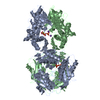 1o90C 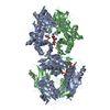 1o92C 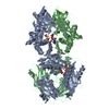 1o93C  1qm4S C: citing same article ( S: Starting model for refinement |
|---|---|
| Similar structure data |

-Links
 PDBj
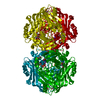
PDBj- Assembly
| Deposited unit | 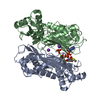
| ||||||||
|---|---|---|---|---|---|---|---|---|---|
| 1 | 
| ||||||||
| Unit cell |
| ||||||||
| Noncrystallographic symmetry (NCS) | NCS oper: (Code: given Matrix: (-0.7849, -0.2307, 0.575), Vector: |
-Components
-Protein , 1 types, 2 molecules AB
| #1: Protein | Mass: 43637.734 Da / Num. of mol.: 2 Source method: isolated from a genetically manipulated source Source: (gene. exp.)  |
|---|
-Non-polymers , 6 types, 185 molecules 


| #2: Chemical | ChemComp-MET /  Type: L-peptide linking / Mass: 149.211 Da / Num. of mol.: 1 / Source method: obtained synthetically / Formula: C5H11NO2S Type: L-peptide linking / Mass: 149.211 Da / Num. of mol.: 1 / Source method: obtained synthetically / Formula: C5H11NO2S | ||||||||
|---|---|---|---|---|---|---|---|---|---|
| #3: Chemical |  Mass: 94.971 Da / Num. of mol.: 2 / Source method: obtained synthetically / Formula: PO4 Mass: 94.971 Da / Num. of mol.: 2 / Source method: obtained synthetically / Formula: PO4#4: Chemical |  Mass: 24.305 Da / Num. of mol.: 3 / Source method: obtained synthetically / Formula: Mg Mass: 24.305 Da / Num. of mol.: 3 / Source method: obtained synthetically / Formula: Mg#5: Chemical |  Mass: 39.098 Da / Num. of mol.: 2 / Source method: obtained synthetically / Formula: K Mass: 39.098 Da / Num. of mol.: 2 / Source method: obtained synthetically / Formula: K#6: Chemical | ChemComp-ATP / |  Mass: 507.181 Da / Num. of mol.: 1 / Source method: obtained synthetically / Formula: C10H16N5O13P3 / Comment: ATP, energy-carrying molecule*YM Mass: 507.181 Da / Num. of mol.: 1 / Source method: obtained synthetically / Formula: C10H16N5O13P3 / Comment: ATP, energy-carrying molecule*YM#7: Water | ChemComp-HOH / | Mass: 18.015 Da / Num. of mol.: 176 / Source method: isolated from a natural source / Formula: H2O |
-Details
| Compound details | CATALYSES THE REACTION BETWEEN ATP AND L-METHIONINE| Sequence details | THE SWISS-PROT ACCESSION FOR THIS ENTRY P13444 HAS A RESIDUE ASN AT POSITION 345. THE DEPOSITORS ...THE SWISS-PROT ACCESSION FOR THIS ENTRY P13444 HAS A RESIDUE ASN AT POSITION 345. THE DEPOSITORS | |
|---|
-Experimental details
-Experiment
| Experiment | Method: X-RAY DIFFRACTION / Number of used crystals: 1 |
|---|
- Sample preparation
| Crystal | Density Matthews: 3.2 Å3/Da / Density % sol: 61 % | |||||||||||||||||||||||||||||||||||||||||||||||||||||||||||||||
|---|---|---|---|---|---|---|---|---|---|---|---|---|---|---|---|---|---|---|---|---|---|---|---|---|---|---|---|---|---|---|---|---|---|---|---|---|---|---|---|---|---|---|---|---|---|---|---|---|---|---|---|---|---|---|---|---|---|---|---|---|---|---|---|---|
| Crystal grow | pH: 7.5 Details: 16% PEG 10000, 10MM L-METHIONINE, 100MM HEPES PH=7.5 (SOAKING IN 30 MM ATP SOLUTION), pH 7.50 | |||||||||||||||||||||||||||||||||||||||||||||||||||||||||||||||
| Crystal grow | *PLUS pH: 8 / Method: vapor diffusion, hanging drop | |||||||||||||||||||||||||||||||||||||||||||||||||||||||||||||||
| Components of the solutions | *PLUS
|
-Data collection
| Diffraction | Mean temperature: 120 K |
|---|---|
| Diffraction source | Source: ROTATING ANODE / Type: ENRAF-NONIUS / Wavelength: 1.5418 |
| Detector | Type: MARRESEARCH / Detector: IMAGE PLATE / Date: Jul 15, 1999 |
| Radiation | Monochromator: GRAPHITE / Protocol: SINGLE WAVELENGTH / Monochromatic (M) / Laue (L): M / Scattering type: x-ray |
| Radiation wavelength | Wavelength: 1.5418 Å / Relative weight: 1 |
| Reflection | Resolution: 2.76→44.7 Å / Num. obs: 27747 / % possible obs: 97.5 % / Observed criterion σ(I): 4 / Redundancy: 4.1 % / Rsym value: 0.074 / Net I/σ(I): 7.7 |
| Reflection shell | Resolution: 2.76→2.92 Å / Redundancy: 4 % / Mean I/σ(I) obs: 1.7 / Rsym value: 0.443 / % possible all: 97.9 |
| Reflection | *PLUS Highest resolution: 2.7 Å / Num. obs: 27790 / Redundancy: 4.1 % / Rmerge(I) obs: 0.078 |
| Reflection shell | *PLUS Highest resolution: 2.7 Å / Lowest resolution: 2.82 Å / % possible obs: 97.5 % / Redundancy: 3.8 % / Rmerge(I) obs: 0.502 / Mean I/σ(I) obs: 1.6 |
- Processing
| Software |
| ||||||||||||||||||||||||||||||||||||||||||||||||||||||||||||
|---|---|---|---|---|---|---|---|---|---|---|---|---|---|---|---|---|---|---|---|---|---|---|---|---|---|---|---|---|---|---|---|---|---|---|---|---|---|---|---|---|---|---|---|---|---|---|---|---|---|---|---|---|---|---|---|---|---|---|---|---|---|
| Refinement | Method to determine structure: MOLECULAR REPLACEMENT Starting model: PDB ENTRY 1QM4 Resolution: 2.9→8 Å / Rfactor Rfree error: 0.008 / Data cutoff high absF: 10000000 / Data cutoff low absF: 0.001 / Isotropic thermal model: RESTRAINED / Cross valid method: THROUGHOUT / σ(F): 3
| ||||||||||||||||||||||||||||||||||||||||||||||||||||||||||||
| Displacement parameters | Biso mean: 37.4 Å2 | ||||||||||||||||||||||||||||||||||||||||||||||||||||||||||||
| Refinement step | Cycle: LAST / Resolution: 2.9→8 Å
| ||||||||||||||||||||||||||||||||||||||||||||||||||||||||||||
| Refine LS restraints |
| ||||||||||||||||||||||||||||||||||||||||||||||||||||||||||||
| LS refinement shell | Resolution: 2.9→3.02 Å / Rfactor Rfree error: 0.028 / Total num. of bins used: 8
| ||||||||||||||||||||||||||||||||||||||||||||||||||||||||||||
| Xplor file | Serial no: 1 / Param file: PARHCSDX.PRO / Topol file: TOPHCSDX.PRO | ||||||||||||||||||||||||||||||||||||||||||||||||||||||||||||
| Refinement | *PLUS Highest resolution: 2.7 Å / Lowest resolution: 25 Å / % reflection Rfree: 7 % / Rfactor Rfree: 0.29 / Rfactor Rwork: 0.24 | ||||||||||||||||||||||||||||||||||||||||||||||||||||||||||||
| Solvent computation | *PLUS | ||||||||||||||||||||||||||||||||||||||||||||||||||||||||||||
| Displacement parameters | *PLUS | ||||||||||||||||||||||||||||||||||||||||||||||||||||||||||||
| Refine LS restraints | *PLUS
|
