 Movie
Movie Controller

[English] 日本語
 Yorodumi
- PDB-6ezn: Cryo-EM structure of the yeast oligosaccharyltransferase (OST) complex -
+ Open data

- Basic information
| Entry | Database: PDB / ID: 6ezn | |||||||||
|---|---|---|---|---|---|---|---|---|---|---|
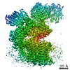
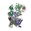
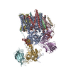
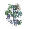
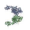
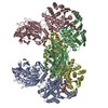
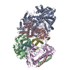
| Title | Cryo-EM structure of the yeast oligosaccharyltransferase (OST) complex | |||||||||
 Components Components | (Dolichyl-diphosphooligosaccharide--protein glycosyltransferase subunit ...) x 8 | |||||||||
 Keywords Keywords | MEMBRANE PROTEIN / OST complex / oligosaccharyltransferase / N-linked glycosylation / yeast | |||||||||
| Function / homology |  Function and homology information Function and homology informationMiscellaneous transport and binding events / dolichol-linked oligosaccharide biosynthetic process / protein O-linked glycosylation via mannose / dolichyl-diphosphooligosaccharide-protein glycotransferase / dolichyl-diphosphooligosaccharide-protein glycotransferase activity / oligosaccharyltransferase complex / : / protein N-linked glycosylation / glycosyltransferase activity / protein-disulfide reductase activity ...Miscellaneous transport and binding events / dolichol-linked oligosaccharide biosynthetic process / protein O-linked glycosylation via mannose / dolichyl-diphosphooligosaccharide-protein glycotransferase / dolichyl-diphosphooligosaccharide-protein glycotransferase activity / oligosaccharyltransferase complex / : / protein N-linked glycosylation / glycosyltransferase activity / protein-disulfide reductase activity / Neutrophil degranulation / post-translational protein modification / nuclear envelope / protein-macromolecule adaptor activity / endoplasmic reticulum membrane / structural molecule activity / endoplasmic reticulum / mitochondrion / membrane / metal ion binding Similarity search - Function | |||||||||
| Biological species |  | |||||||||
| Method | ELECTRON MICROSCOPY / single particle reconstruction / cryo EM / Resolution: 3.3 Å | |||||||||
 Authors Authors | Wild, R. / Kowal, J. / Eyring, J. / Ngwa, E.M. / Aebi, M. / Locher, K.P. | |||||||||
| Funding support |  Switzerland, 2items Switzerland, 2items
| |||||||||
 Citation Citation | Journal: Science / Year: 2018 Title: Structure of the yeast oligosaccharyltransferase complex gives insight into eukaryotic N-glycosylation. Authors: Rebekka Wild / Julia Kowal / Jillianne Eyring / Elsy M Ngwa / Markus Aebi / Kaspar P Locher / Abstract: Oligosaccharyltransferase (OST) is an essential membrane protein complex in the endoplasmic reticulum, where it transfers an oligosaccharide from a dolichol-pyrophosphate-activated donor to ...Oligosaccharyltransferase (OST) is an essential membrane protein complex in the endoplasmic reticulum, where it transfers an oligosaccharide from a dolichol-pyrophosphate-activated donor to glycosylation sites of secretory proteins. Here we describe the atomic structure of yeast OST determined by cryo-electron microscopy, revealing a conserved subunit arrangement. The active site of the catalytic STT3 subunit points away from the center of the complex, allowing unhindered access to substrates. The dolichol-pyrophosphate moiety binds to a lipid-exposed groove of STT3, whereas two noncatalytic subunits and an ordered N-glycan form a membrane-proximal pocket for the oligosaccharide. The acceptor polypeptide site faces an oxidoreductase domain in stand-alone OST complexes or is immediately adjacent to the translocon, suggesting how eukaryotic OSTs efficiently glycosylate a large number of polypeptides before their folding. | |||||||||
| History |
|
- Structure visualization
| Movie |
Movie viewer |
|---|---|
| Structure viewer | Molecule: MolmilJmol/JSmol |
- Downloads & links
-Download
| PDBx/mmCIF format | 6ezn.cif.gz | 399 KB | Display | PDBx/mmCIF format |
|---|---|---|---|---|
| PDB format | pdb6ezn.ent.gz | 314.3 KB | Display | PDB format |
| PDBx/mmJSON format | 6ezn.json.gz | Tree view | PDBx/mmJSON format | |
| Others |  Other downloads Other downloads |
-Validation report
| Arichive directory | https://data.pdbj.org/pub/pdb/validation_reports/ez/6eznftp://data.pdbj.org/pub/pdb/validation_reports/ez/6ezn | HTTPS FTP |
|---|
-Related structure data
| Related structure data |  4161MC 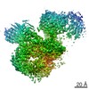 4257C M: map data used to model this data C: citing same article ( |
|---|---|
| Similar structure data |

-Links
 PDBj
PDBj


- Assembly
| Deposited unit | 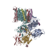
|
|---|---|
| 1 |
|
-Components
-Dolichyl-diphosphooligosaccharide--protein glycosyltransferase subunit ... , 8 types, 8 molecules ABCDEFGH
| #1: Protein | Mass: 54116.477 Da / Num. of mol.: 1 / Source method: isolated from a natural source Source: (natural) Strain: ATCC 204508 / S288c References: UniProt: P41543, dolichyl-diphosphooligosaccharide-protein glycotransferase |
|---|---|
| #2: Protein | Mass: 14712.531 Da / Num. of mol.: 1 / Source method: isolated from a natural source Source: (natural) Strain: ATCC 204508 / S288c References: UniProt: P46964, dolichyl-diphosphooligosaccharide-protein glycotransferase |
| #3: Protein | Mass: 39518.160 Da / Num. of mol.: 1 / Source method: isolated from a natural source Source: (natural) Strain: ATCC 204508 / S288c References: UniProt: P48439, dolichyl-diphosphooligosaccharide-protein glycotransferase |
| #4: Protein/peptide | Mass: 3986.696 Da / Num. of mol.: 1 / Source method: isolated from a natural source Source: (natural) Strain: ATCC 204508 / S288c References: UniProt: Q99380, dolichyl-diphosphooligosaccharide-protein glycotransferase |
| #5: Protein | Mass: 9525.090 Da / Num. of mol.: 1 / Source method: isolated from a natural source Source: (natural) Strain: ATCC 204508 / S288c References: UniProt: Q92316, dolichyl-diphosphooligosaccharide-protein glycotransferase |
| #6: Protein | Mass: 81604.539 Da / Num. of mol.: 1 / Source method: isolated from a natural source Source: (natural) Strain: ATCC 204508 / S288c References: UniProt: P39007, dolichyl-diphosphooligosaccharide-protein glycotransferase |
| #7: Protein | Mass: 49444.438 Da / Num. of mol.: 1 / Source method: isolated from a natural source Source: (natural) Strain: ATCC 204508 / S288c References: UniProt: P33767, dolichyl-diphosphooligosaccharide-protein glycotransferase |
| #8: Protein | Mass: 31682.832 Da / Num. of mol.: 1 / Source method: isolated from a natural source Source: (natural) Strain: ATCC 204508 / S288c References: UniProt: Q02795, dolichyl-diphosphooligosaccharide-protein glycotransferase |
-Sugars , 4 types, 5 molecules 
| #9: Polysaccharide | beta-D-mannopyranose-(1-4)-2-acetamido-2-deoxy-beta-D-glucopyranose-(1-4)-2-acetamido-2-deoxy-beta- ...beta-D-mannopyranose-(1-4)-2-acetamido-2-deoxy-beta-D-glucopyranose-(1-4)-2-acetamido-2-deoxy-beta-D-glucopyranose Source method: isolated from a genetically manipulated source | ||||
|---|---|---|---|---|---|
| #10: Polysaccharide | Source method: isolated from a genetically manipulated source #11: Polysaccharide | alpha-D-mannopyranose-(1-2)-alpha-D-mannopyranose-(1-2)-alpha-D-mannopyranose-(1-3)-[alpha-D- ...alpha-D-mannopyranose-(1-2)-alpha-D-mannopyranose-(1-2)-alpha-D-mannopyranose-(1-3)-[alpha-D-mannopyranose-(1-3)-alpha-D-mannopyranose-(1-6)]beta-D-mannopyranose-(1-4)-2-acetamido-2-deoxy-beta-D-glucopyranose-(1-4)-2-acetamido-2-deoxy-beta-D-glucopyranose | Source method: isolated from a genetically manipulated source #14: Sugar | ChemComp-NAG / |  Type: D-saccharide, beta linking / Mass: 221.208 Da / Num. of mol.: 1 Type: D-saccharide, beta linking / Mass: 221.208 Da / Num. of mol.: 1Source method: isolated from a genetically manipulated source Formula: C8H15NO6 |
-Non-polymers , 2 types, 5 molecules 

| #12: Chemical | ChemComp-CPL /  Mass: 758.060 Da / Num. of mol.: 4 / Source method: obtained synthetically / Formula: C42H80NO8P / Comment: phospholipid*YM Mass: 758.060 Da / Num. of mol.: 4 / Source method: obtained synthetically / Formula: C42H80NO8P / Comment: phospholipid*YM#13: Chemical | ChemComp-PTY / |  Mass: 734.039 Da / Num. of mol.: 1 / Source method: obtained synthetically / Formula: C40H80NO8P / Comment: phospholipid*YM Mass: 734.039 Da / Num. of mol.: 1 / Source method: obtained synthetically / Formula: C40H80NO8P / Comment: phospholipid*YM |
|---|
-Details
| Compound details | Amino acid residues H22 to H65 in Swp1 were built as PolyA chain and renamed to specific amino ...Amino acid residues H22 to H65 in Swp1 were built as PolyA chain and renamed to specific amino acids according to residue numbers on request by wwPDB |
|---|---|
| Has protein modification | Y |
-Experimental details
-Experiment
| Experiment | Method: ELECTRON MICROSCOPY |
|---|---|
| EM experiment | Aggregation state: PARTICLE / 3D reconstruction method: single particle reconstruction |
- Sample preparation
| Component | Name: yeast oligosaccharyltransferase (OST) complex / Type: COMPLEX / Entity ID: #1-#8 / Source: NATURAL |
|---|---|
| Source (natural) | Organism: |
| Buffer solution | pH: 7.5 / Details: 20 mM HEPES, pH 7.5, 150 mM NaCl |
| Specimen | Conc.: 0.5 mg/ml / Embedding applied: NO / Shadowing applied: NO / Staining applied: NO / Vitrification applied: YES / Details: OST complex reconstituted into nanodisc |
| Specimen support | Grid material: COPPER / Grid mesh size: 300 divisions/in. / Grid type: Quantifoil R1.2/1.3 |
| Vitrification | Instrument: FEI VITROBOT MARK IV / Cryogen name: ETHANE-PROPANE / Humidity: 95 % / Chamber temperature: 277 K Details: Quantifoil carbon grids (300 mesh, copper) were glow discharged at 25 mA for 40 s. 3.5 ul of nanodisc-reconstituted OST sample at concentration of 0.5 mg/ml was applied onto the grid and ...Details: Quantifoil carbon grids (300 mesh, copper) were glow discharged at 25 mA for 40 s. 3.5 ul of nanodisc-reconstituted OST sample at concentration of 0.5 mg/ml was applied onto the grid and grids were blotted for 3 s and flash-frozen in a mixture of liquid ethane and propane cooled by liquid nitrogen. |
- Electron microscopy imaging
| Experimental equipment |  Model: Titan Krios / Image courtesy: FEI Company |
|---|---|
| Microscopy | Model: FEI TITAN KRIOS |
| Electron gun | Electron source:  FIELD EMISSION GUN / Accelerating voltage: 300 kV / Illumination mode: FLOOD BEAM FIELD EMISSION GUN / Accelerating voltage: 300 kV / Illumination mode: FLOOD BEAM |
| Electron lens | Mode: BRIGHT FIELD / Cs: 2.7 mm / C2 aperture diameter: 100 µm / Alignment procedure: COMA FREE |
| Specimen holder | Cryogen: NITROGEN / Specimen holder model: FEI TITAN KRIOS AUTOGRID HOLDER |
| Image recording | Average exposure time: 0.25 sec. / Electron dose: 2 e/Å2 / Film or detector model: GATAN K2 SUMMIT (4k x 4k) / Num. of grids imaged: 2 / Num. of real images: 3915 |
| EM imaging optics | Energyfilter name: GIF Quantum LS |
- Processing
| Software | Name: PHENIX / Version: 1.11.1_2575: / Classification: refinement | ||||||||||||||||||||||||||||||||
|---|---|---|---|---|---|---|---|---|---|---|---|---|---|---|---|---|---|---|---|---|---|---|---|---|---|---|---|---|---|---|---|---|---|
| EM software |
| ||||||||||||||||||||||||||||||||
| CTF correction | Type: PHASE FLIPPING AND AMPLITUDE CORRECTION | ||||||||||||||||||||||||||||||||
| Particle selection | Num. of particles selected: 486361 | ||||||||||||||||||||||||||||||||
| Symmetry | Point symmetry: C1 (asymmetric) | ||||||||||||||||||||||||||||||||
| 3D reconstruction | Resolution: 3.3 Å / Resolution method: FSC 0.143 CUT-OFF / Num. of particles: 110091 / Symmetry type: POINT | ||||||||||||||||||||||||||||||||
| Atomic model building | Protocol: AB INITIO MODEL | ||||||||||||||||||||||||||||||||
| Refine LS restraints |
|
