 Movie
Movie Controller


+ Open data

- Basic information
| Entry | Database: PDB / ID: 7btp | ||||||
|---|---|---|---|---|---|---|---|
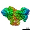
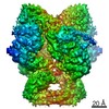
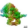
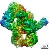
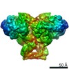
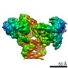
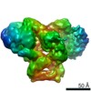
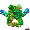
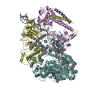
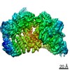
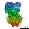
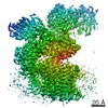
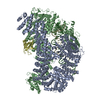
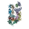
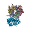
| Title | EcoR124I-Ocr in Restriction-Alleviation State | ||||||
 Components Components |
| ||||||
 Keywords Keywords | IMMUNE SYSTEM / Cryoelectron microscopy / Innate immune mechanism / Complex | ||||||
| Function / homology |  Function and homology information Function and homology informationtype I site-specific deoxyribonuclease / type I site-specific deoxyribonuclease activity / symbiont-mediated evasion of host restriction-modification system / N-methyltransferase activity / site-specific DNA-methyltransferase (adenine-specific) / site-specific DNA-methyltransferase (adenine-specific) activity / DNA restriction-modification system / methylation / symbiont-mediated suppression of host innate immune response / DNA binding / ATP binding Similarity search - Function | ||||||
| Biological species |    Escherichia phage T7 (virus) Escherichia phage T7 (virus) | ||||||
| Method | ELECTRON MICROSCOPY / single particle reconstruction / cryo EM / Resolution: 4.01 Å | ||||||
 Authors Authors | Gao, Y. / Gao, P. | ||||||
 Citation Citation | Journal: Nat Microbiol / Year: 2020 Title: Structural insights into assembly, operation and inhibition of a type I restriction-modification system. Authors: Yina Gao / Duanfang Cao / Jingpeng Zhu / Han Feng / Xiu Luo / Songqing Liu / Xiao-Xue Yan / Xinzheng Zhang / Pu Gao /  Abstract: Type I restriction-modification (R-M) systems are widespread in prokaryotic genomes and provide robust protection against foreign DNA. They are multisubunit enzymes with methyltransferase, ...Type I restriction-modification (R-M) systems are widespread in prokaryotic genomes and provide robust protection against foreign DNA. They are multisubunit enzymes with methyltransferase, endonuclease and translocase activities. Despite extensive studies over the past five decades, little is known about the molecular mechanisms of these sophisticated machines. Here, we report the cryo-electron microscopy structures of the representative EcoR124I R-M system in different assemblies (RMS, RMS and MS) bound to target DNA and the phage and mobile genetic element-encoded anti-restriction proteins Ocr and ArdA. EcoR124I can precisely regulate different enzymatic activities by adopting distinct conformations. The marked conformational transitions of EcoR124I are dependent on the intrinsic flexibility at both the individual-subunit and assembled-complex levels. Moreover, Ocr and ArdA use a DNA-mimicry strategy to inhibit multiple activities, but do not block the conformational transitions of the complexes. These structural findings, complemented by mutational studies of key intermolecular contacts, provide insights into assembly, operation and inhibition mechanisms of type I R-M systems. | ||||||
| History |
|
- Structure visualization
| Movie |
Movie viewer |
|---|---|
| Structure viewer | Molecule: MolmilJmol/JSmol |
- Downloads & links
-Download
| PDBx/mmCIF format | 7btp.cif.gz | 518.5 KB | Display | PDBx/mmCIF format |
|---|---|---|---|---|
| PDB format | pdb7btp.ent.gz | 406.3 KB | Display | PDB format |
| PDBx/mmJSON format | 7btp.json.gz | Tree view | PDBx/mmJSON format | |
| Others |  Other downloads Other downloads |
-Validation report
| Arichive directory | https://data.pdbj.org/pub/pdb/validation_reports/bt/7btpftp://data.pdbj.org/pub/pdb/validation_reports/bt/7btp | HTTPS FTP |
|---|
-Related structure data
| Related structure data | 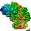 30181MC 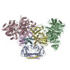 7bstC 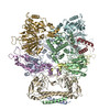 7btoC 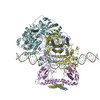 7btqC 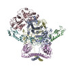 7btrC M: map data used to model this data C: citing same article ( |
|---|---|
| Similar structure data |


-Links
 PDBj
PDBj- Assembly
| Deposited unit | 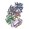
|
|---|---|
| 1 |
|
-Components
| #1: Protein | Mass: 46235.773 Da / Num. of mol.: 1 Source method: isolated from a genetically manipulated source Source: (gene. exp.) | ||
|---|---|---|---|
| #2: Protein | Mass: 120278.859 Da / Num. of mol.: 1 Source method: isolated from a genetically manipulated source Source: (gene. exp.) References: UniProt: Q304R3, UniProt: P10486*PLUS, type I site-specific deoxyribonuclease | ||
| #3: Protein | Mass: 58077.090 Da / Num. of mol.: 2 Source method: isolated from a genetically manipulated source Source: (gene. exp.) References: UniProt: P10484, site-specific DNA-methyltransferase (adenine-specific) #4: Protein | Mass: 13819.015 Da / Num. of mol.: 2 Source method: isolated from a genetically manipulated source Source: (gene. exp.) Escherichia phage T7 (virus) / Gene: 0.3 / Production host: |
-Experimental details
-Experiment
| Experiment | Method: ELECTRON MICROSCOPY |
|---|---|
| EM experiment | Aggregation state: PARTICLE / 3D reconstruction method: single particle reconstruction |
- Sample preparation
| Component | Name: EcoR124I-Ocr / Type: COMPLEX / Entity ID: all / Source: RECOMBINANT |
|---|---|
| Source (natural) | Organism: |
| Source (recombinant) | Organism: |
| Buffer solution | pH: 7.5 |
| Specimen | Embedding applied: NO / Shadowing applied: NO / Staining applied: NO / Vitrification applied: YES |
| Vitrification | Cryogen name: ETHANE |
- Electron microscopy imaging
| Experimental equipment |  Model: Titan Krios / Image courtesy: FEI Company |
|---|---|
| Microscopy | Model: FEI TITAN KRIOS |
| Electron gun | Electron source:  FIELD EMISSION GUN / Accelerating voltage: 300 kV / Illumination mode: FLOOD BEAM FIELD EMISSION GUN / Accelerating voltage: 300 kV / Illumination mode: FLOOD BEAM |
| Electron lens | Mode: BRIGHT FIELD |
| Image recording | Electron dose: 60 e/Å2 / Film or detector model: GATAN K2 SUMMIT (4k x 4k) |
- Processing
| CTF correction | Type: PHASE FLIPPING AND AMPLITUDE CORRECTION |
|---|---|
| 3D reconstruction | Resolution: 4.01 Å / Resolution method: FSC 0.143 CUT-OFF / Num. of particles: 197498 / Symmetry type: POINT |
