 Movie
Movie Controller

[English] 日本語
 Yorodumi
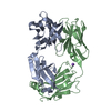
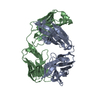
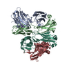
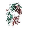
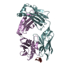
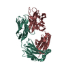
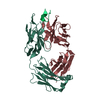
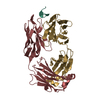
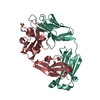
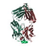
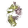
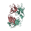
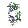
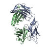
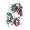
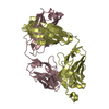
- PDB-6elj: FAB Fragment. AbVance: Increasing our knowledge of antibody struc... -
+ Open data

- Basic information
| Entry | Database: PDB / ID: 6elj | ||||||
|---|---|---|---|---|---|---|---|
| Title | FAB Fragment. AbVance: Increasing our knowledge of antibody structural space to enable faster and better decision making in antibody drug discovery | ||||||
 Components Components |
| ||||||
 Keywords Keywords | IMMUNE SYSTEM / ANTIBODY / fAB FRAGMENT / AbVance project / Pistoia Alliance | ||||||
| Function / homology | Immunoglobulins / Immunoglobulin-like / Sandwich / Mainly Beta Function and homology information Function and homology information | ||||||
| Biological species |  Homo sapiens (human) Homo sapiens (human) | ||||||
| Method |  X-RAY DIFFRACTION / SYNCHROTRON / MOLECULAR REPLACEMENT / Resolution: 1.9 Å X-RAY DIFFRACTION / SYNCHROTRON / MOLECULAR REPLACEMENT / Resolution: 1.9 Å | ||||||
 Authors Authors | Benz, J. / Weigand, S. / Dengl, S. / Schlothauer, T. / Auer, J. / Ehler, A. / Kettenberger, H. / Lorenz, S. / Hirschheydt, T. / Georges, G. | ||||||
 Citation Citation | Journal: To Be Published Title: AbVance: increasing our knowledge of antibody structural space to enable faster and better decision making in antibody drug discovery Authors: Benz, J. / Georges, G. | ||||||
| History |
|
- Structure visualization
| Structure viewer | Molecule: MolmilJmol/JSmol |
|---|
- Downloads & links
-Download
| PDBx/mmCIF format | 6elj.cif.gz | 344.5 KB | Display | PDBx/mmCIF format |
|---|---|---|---|---|
| PDB format | pdb6elj.ent.gz | 281.6 KB | Display | PDB format |
| PDBx/mmJSON format | 6elj.json.gz | Tree view | PDBx/mmJSON format | |
| Others |  Other downloads Other downloads |
-Validation report
| Arichive directory | https://data.pdbj.org/pub/pdb/validation_reports/el/6eljftp://data.pdbj.org/pub/pdb/validation_reports/el/6elj | HTTPS FTP |
|---|
-Related structure data


-Links
 PDBj
PDBj

- Assembly
| Deposited unit | 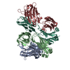
| ||||||||
|---|---|---|---|---|---|---|---|---|---|
| 1 | 
| ||||||||
| 2 | 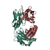
| ||||||||
| Unit cell |
|
-Components
| #1: Antibody | Mass: 24570.447 Da / Num. of mol.: 2 Source method: isolated from a genetically manipulated source Source: (gene. exp.) Homo sapiens (human) / Cell line (production host): HEK293 / Production host: Homo sapiens (human)#2: Antibody | Mass: 23552.129 Da / Num. of mol.: 2 Source method: isolated from a genetically manipulated source Source: (gene. exp.) Homo sapiens (human) / Cell line (production host): HEK293 / Production host: Homo sapiens (human)#3: Water | ChemComp-HOH / |  Mass: 18.015 Da / Num. of mol.: 379 / Source method: isolated from a natural source / Formula: H2O Mass: 18.015 Da / Num. of mol.: 379 / Source method: isolated from a natural source / Formula: H2OHas protein modification | Y | |
|---|
-Experimental details
-Experiment
| Experiment | Method: X-RAY DIFFRACTION / Number of used crystals: 1 |
|---|
- Sample preparation
| Crystal | Density Matthews: 2.37 Å3/Da / Density % sol: 48 % |
|---|---|
| Crystal grow | Temperature: 293 K / Method: vapor diffusion, sitting drop / Details: 0.1 M Na cacodylat 6.5 pH 25 %w/v PEG 4K |
-Data collection
| Diffraction | Mean temperature: 100 K |
|---|---|
| Diffraction source | Source: SYNCHROTRON / Site: SLS  / Beamline: X10SA / Wavelength: 1 Å / Beamline: X10SA / Wavelength: 1 Å |
| Detector | Type: DECTRIS PILATUS3 6M / Detector: PIXEL / Date: Mar 21, 2014 |
| Radiation | Protocol: SINGLE WAVELENGTH / Monochromatic (M) / Laue (L): M / Scattering type: x-ray |
| Radiation wavelength | Wavelength: 1 Å / Relative weight: 1 |
| Reflection | Resolution: 1.9→44.24 Å / Num. obs: 67823 / % possible obs: 97.6 % / Redundancy: 3.42 % / Biso Wilson estimate: 31.03 Å2 / Rmerge(I) obs: 0.01 / Rsym value: 0.09 / Net I/σ(I): 7.55 |
| Reflection shell | Resolution: 1.9→2 Å / Rsym value: 0.68 / % possible all: 96.2 |
- Processing
| Software |
| |||||||||||||||||||||||||||||||||||||||||||||||||||||||||||||||||||||||||||||||||||||||||||||||||||||||||||||||||||||||||||||
|---|---|---|---|---|---|---|---|---|---|---|---|---|---|---|---|---|---|---|---|---|---|---|---|---|---|---|---|---|---|---|---|---|---|---|---|---|---|---|---|---|---|---|---|---|---|---|---|---|---|---|---|---|---|---|---|---|---|---|---|---|---|---|---|---|---|---|---|---|---|---|---|---|---|---|---|---|---|---|---|---|---|---|---|---|---|---|---|---|---|---|---|---|---|---|---|---|---|---|---|---|---|---|---|---|---|---|---|---|---|---|---|---|---|---|---|---|---|---|---|---|---|---|---|---|---|---|
| Refinement | Method to determine structure: MOLECULAR REPLACEMENT Starting model: in house Fab structure Resolution: 1.9→44.24 Å / Cor.coef. Fo:Fc: 0.955 / Cor.coef. Fo:Fc free: 0.934 / SU R Cruickshank DPI: 0.146 / Cross valid method: THROUGHOUT / σ(F): 0 / SU R Blow DPI: 0.143 / SU Rfree Blow DPI: 0.135 / SU Rfree Cruickshank DPI: 0.137
| |||||||||||||||||||||||||||||||||||||||||||||||||||||||||||||||||||||||||||||||||||||||||||||||||||||||||||||||||||||||||||||
| Displacement parameters | Biso mean: 35.97 Å2
| |||||||||||||||||||||||||||||||||||||||||||||||||||||||||||||||||||||||||||||||||||||||||||||||||||||||||||||||||||||||||||||
| Refine analyze | Luzzati coordinate error obs: 0.24 Å | |||||||||||||||||||||||||||||||||||||||||||||||||||||||||||||||||||||||||||||||||||||||||||||||||||||||||||||||||||||||||||||
| Refinement step | Cycle: 1 / Resolution: 1.9→44.24 Å
| |||||||||||||||||||||||||||||||||||||||||||||||||||||||||||||||||||||||||||||||||||||||||||||||||||||||||||||||||||||||||||||
| Refine LS restraints |
| |||||||||||||||||||||||||||||||||||||||||||||||||||||||||||||||||||||||||||||||||||||||||||||||||||||||||||||||||||||||||||||
| LS refinement shell | Resolution: 1.9→1.95 Å / Total num. of bins used: 20
| |||||||||||||||||||||||||||||||||||||||||||||||||||||||||||||||||||||||||||||||||||||||||||||||||||||||||||||||||||||||||||||
| Refinement TLS params. | Method: refined / Refine-ID: X-RAY DIFFRACTION
| |||||||||||||||||||||||||||||||||||||||||||||||||||||||||||||||||||||||||||||||||||||||||||||||||||||||||||||||||||||||||||||
| Refinement TLS group |
|
