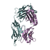
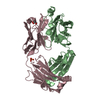
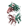
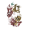
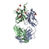
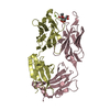
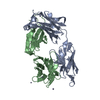
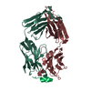
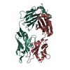
Journal: Nat Struct Mol Biol / Year: 2013 Title: Supersite of immune vulnerability on the glycosylated face of HIV-1 envelope glycoprotein gp120. Authors: Leopold Kong / Jeong Hyun Lee / Katie J Doores / Charles D Murin / Jean-Philippe Julien / Ryan McBride / Yan Liu / Andre Marozsan / Albert Cupo / Per-Johan Klasse / Simon Hoffenberg / ...Authors: Leopold Kong / Jeong Hyun Lee / Katie J Doores / Charles D Murin / Jean-Philippe Julien / Ryan McBride / Yan Liu / Andre Marozsan / Albert Cupo / Per-Johan Klasse / Simon Hoffenberg / Michael Caulfield / C Richter King / Yuanzi Hua / Khoa M Le / Reza Khayat / Marc C Deller / Thomas Clayton / Henry Tien / Ten Feizi / Rogier W Sanders / James C Paulson / John P Moore / Robyn L Stanfield / Dennis R Burton / Andrew B Ward / Ian A Wilson / Abstract: A substantial proportion of the broadly neutralizing antibodies (bnAbs) identified in certain HIV-infected donors recognize glycan-dependent epitopes on HIV-1 gp120. Here we elucidate how the bnAb ...A substantial proportion of the broadly neutralizing antibodies (bnAbs) identified in certain HIV-infected donors recognize glycan-dependent epitopes on HIV-1 gp120. Here we elucidate how the bnAb PGT 135 binds its Asn332 glycan-dependent epitope from its 3.1-Å crystal structure with gp120, CD4 and Fab 17b. PGT 135 interacts with glycans at Asn332, Asn392 and Asn386, using long CDR loops H1 and H3 to penetrate the glycan shield and access the gp120 protein surface. EM reveals that PGT 135 can accommodate the conformational and chemical diversity of gp120 glycans by altering its angle of engagement. Combined structural studies of PGT 135, PGT 128 and 2G12 show that this Asn332-dependent antigenic region is highly accessible and much more extensive than initially appreciated, which allows for multiple binding modes and varied angles of approach; thereby it represents a supersite of vulnerability for antibody neutralization.
Mass: 25575.814 Da / Num. of mol.: 1 / Fragment: Fab Source method: isolated from a genetically manipulated source Source: (gene. exp.) Homo sapiens (human) / Production host: Spodoptera frugiperda (fall armyworm)
#2: Antibody
PGT135LightChain
Mass: 23795.467 Da / Num. of mol.: 1 / Fragment: Fab Source method: isolated from a genetically manipulated source Source: (gene. exp.) Homo sapiens (human) / Production host: Spodoptera frugiperda (fall armyworm)
In the structure databanks used in Yorodumi, some data are registered as the other names, "COVID-19 virus" and "2019-nCoV". Here are the details of the virus and the list of structure data.
Jan 31, 2019. EMDB accession codes are about to change! (news from PDBe EMDB page)
EMDB accession codes are about to change! (news from PDBe EMDB page)
The allocation of 4 digits for EMDB accession codes will soon come to an end. Whilst these codes will remain in use, new EMDB accession codes will include an additional digit and will expand incrementally as the available range of codes is exhausted. The current 4-digit format prefixed with “EMD-” (i.e. EMD-XXXX) will advance to a 5-digit format (i.e. EMD-XXXXX), and so on. It is currently estimated that the 4-digit codes will be depleted around Spring 2019, at which point the 5-digit format will come into force.
The EM Navigator/Yorodumi systems omit the EMD- prefix.
Related info.:Q: What is EMD? / ID/Accession-code notation in Yorodumi/EM Navigator
Yorodumi is a browser for structure data from EMDB, PDB, SASBDB, etc.
This page is also the successor to EM Navigator detail page, and also detail information page/front-end page for Omokage search.
The word "yorodu" (or yorozu) is an old Japanese word meaning "ten thousand". "mi" (miru) is to see.
Related info.:EMDB / PDB / SASBDB / Comparison of 3 databanks / Yorodumi Search / Aug 31, 2016. New EM Navigator & Yorodumi / Yorodumi Papers / Jmol/JSmol / Function and homology information / Changes in new EM Navigator and Yorodumi
 Movie
Movie Controller


 Open data
 Basic information
 Components
 Keywords
 Function and homology information
 Homo sapiens (human)
 X-RAY DIFFRACTION /
  Authors
 Citation
 Structure visualization
 Downloads & links
 Other downloads
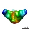


 PDBj
PDBj

 Assembly
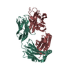

 Spodoptera frugiperda (fall armyworm)
 Mass: 18.015 Da / Num. of mol.: 273 / Source method: isolated from a natural source / Formula: H2O
 Sample preparation
 Processing