 Movie
Movie Controller

[English] 日本語
 Yorodumi
- PDB-4jm2: Crystal Structure of PGT 135 Fab in Complex with gp120 Core Prote... -
+ Open data

- Basic information
| Entry | Database: PDB / ID: 4jm2 | |||||||||
|---|---|---|---|---|---|---|---|---|---|---|
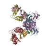
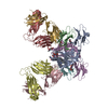
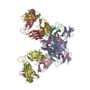
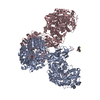
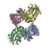
| Title | Crystal Structure of PGT 135 Fab in Complex with gp120 Core Protein from HIV-1 Strain JR-FL Bound to CD4 and 17b Fab | |||||||||
 Components Components |
| |||||||||
 Keywords Keywords | IMMUNE SYSTEM/VIRAL PROTEIN / Immunoglobulin Fold / IMMUNE SYSTEM-VIRAL PROTEIN complex | |||||||||
| Function / homology |  Function and homology information Function and homology informationhelper T cell enhancement of adaptive immune response / interleukin-16 binding / interleukin-16 receptor activity / cellular response to ionomycin / maintenance of protein location in cell / response to methamphetamine hydrochloride / T cell selection / MHC class II protein binding / interleukin-15-mediated signaling pathway / cellular response to granulocyte macrophage colony-stimulating factor stimulus ...helper T cell enhancement of adaptive immune response / interleukin-16 binding / interleukin-16 receptor activity / cellular response to ionomycin / maintenance of protein location in cell / response to methamphetamine hydrochloride / T cell selection / MHC class II protein binding / interleukin-15-mediated signaling pathway / cellular response to granulocyte macrophage colony-stimulating factor stimulus / positive regulation of monocyte differentiation / Alpha-defensins / Nef Mediated CD4 Down-regulation / regulation of T cell activation / response to vitamin D / Other interleukin signaling / leukocyte chemotaxis / extracellular matrix structural constituent / T cell receptor complex / enzyme-linked receptor protein signaling pathway / Translocation of ZAP-70 to Immunological synapse / Phosphorylation of CD3 and TCR zeta chains / macrophage differentiation / Generation of second messenger molecules / immunoglobulin binding / T cell differentiation / Co-inhibition by PD-1 / positive regulation of calcium ion transport into cytosol / Binding and entry of HIV virion / coreceptor activity / symbiont-mediated perturbation of host defense response / positive regulation of plasma membrane raft polarization / positive regulation of receptor clustering / positive regulation of interleukin-2 production / positive regulation of T cell proliferation / cell surface receptor protein tyrosine kinase signaling pathway / protein tyrosine kinase binding / host cell endosome membrane / T cell activation / clathrin-coated endocytic vesicle membrane / Vpu mediated degradation of CD4 / positive regulation of inflammatory response / MHC class II protein complex binding / response to estradiol / transmembrane signaling receptor activity / T cell receptor signaling pathway / Downstream TCR signaling / Cargo recognition for clathrin-mediated endocytosis / Clathrin-mediated endocytosis / virus receptor activity / signaling receptor activity / clathrin-dependent endocytosis of virus by host cell / response to ethanol / adaptive immune response / early endosome / cell surface receptor signaling pathway / cell adhesion / immune response / viral protein processing / membrane raft / endoplasmic reticulum lumen / fusion of virus membrane with host plasma membrane / external side of plasma membrane / signaling receptor binding / fusion of virus membrane with host endosome membrane / viral envelope / symbiont entry into host cell / lipid binding / protein kinase binding / endoplasmic reticulum membrane / virion attachment to host cell / host cell plasma membrane / virion membrane / structural molecule activity / enzyme binding / signal transduction / protein homodimerization activity / : / zinc ion binding / membrane / identical protein binding / plasma membrane Similarity search - Function | |||||||||
| Biological species |  Homo sapiens (human) Homo sapiens (human)  Human immunodeficiency virus 1 Human immunodeficiency virus 1 | |||||||||
| Method |  X-RAY DIFFRACTION / SYNCHROTRON / MOLECULAR REPLACEMENT / Resolution: 3.1 Å X-RAY DIFFRACTION / SYNCHROTRON / MOLECULAR REPLACEMENT / Resolution: 3.1 Å | |||||||||
 Authors Authors | Kong, L. / Wilson, I.A. | |||||||||
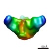
 Citation Citation | Journal: Nat Struct Mol Biol / Year: 2013 Title: Supersite of immune vulnerability on the glycosylated face of HIV-1 envelope glycoprotein gp120. Authors: Leopold Kong / Jeong Hyun Lee / Katie J Doores / Charles D Murin / Jean-Philippe Julien / Ryan McBride / Yan Liu / Andre Marozsan / Albert Cupo / Per-Johan Klasse / Simon Hoffenberg / ...Authors: Leopold Kong / Jeong Hyun Lee / Katie J Doores / Charles D Murin / Jean-Philippe Julien / Ryan McBride / Yan Liu / Andre Marozsan / Albert Cupo / Per-Johan Klasse / Simon Hoffenberg / Michael Caulfield / C Richter King / Yuanzi Hua / Khoa M Le / Reza Khayat / Marc C Deller / Thomas Clayton / Henry Tien / Ten Feizi / Rogier W Sanders / James C Paulson / John P Moore / Robyn L Stanfield / Dennis R Burton / Andrew B Ward / Ian A Wilson /  Abstract: A substantial proportion of the broadly neutralizing antibodies (bnAbs) identified in certain HIV-infected donors recognize glycan-dependent epitopes on HIV-1 gp120. Here we elucidate how the bnAb ...A substantial proportion of the broadly neutralizing antibodies (bnAbs) identified in certain HIV-infected donors recognize glycan-dependent epitopes on HIV-1 gp120. Here we elucidate how the bnAb PGT 135 binds its Asn332 glycan-dependent epitope from its 3.1-Å crystal structure with gp120, CD4 and Fab 17b. PGT 135 interacts with glycans at Asn332, Asn392 and Asn386, using long CDR loops H1 and H3 to penetrate the glycan shield and access the gp120 protein surface. EM reveals that PGT 135 can accommodate the conformational and chemical diversity of gp120 glycans by altering its angle of engagement. Combined structural studies of PGT 135, PGT 128 and 2G12 show that this Asn332-dependent antigenic region is highly accessible and much more extensive than initially appreciated, which allows for multiple binding modes and varied angles of approach; thereby it represents a supersite of vulnerability for antibody neutralization. | |||||||||
| History |
|
- Structure visualization
| Structure viewer | Molecule: MolmilJmol/JSmol |
|---|
- Downloads & links
-Download
| PDBx/mmCIF format | 4jm2.cif.gz | 290.7 KB | Display | PDBx/mmCIF format |
|---|---|---|---|---|
| PDB format | pdb4jm2.ent.gz | 230.7 KB | Display | PDB format |
| PDBx/mmJSON format | 4jm2.json.gz | Tree view | PDBx/mmJSON format | |
| Others |  Other downloads Other downloads |
-Validation report
| Arichive directory | https://data.pdbj.org/pub/pdb/validation_reports/jm/4jm2ftp://data.pdbj.org/pub/pdb/validation_reports/jm/4jm2 | HTTPS FTP |
|---|
-Related structure data


-Links
 PDBj
PDBj


- Assembly
| Deposited unit | 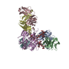
| ||||||||
|---|---|---|---|---|---|---|---|---|---|
| 1 |
| ||||||||
| Unit cell |
|
-Components
-Protein , 2 types, 2 molecules EF
| #5: Protein | Mass: 35991.883 Da / Num. of mol.: 1 / Fragment: core with mini V3 loop Source method: isolated from a genetically manipulated source Source: (gene. exp.) Human immunodeficiency virus 1 / Strain: JRFL / Cell line (production host): HEK 293S / Production host: Homo sapiens (human) / References: UniProt: Q75760*PLUS |
|---|---|
| #6: Protein | Mass: 20503.260 Da / Num. of mol.: 1 Fragment: Ig-like V-type and C2-type 1 domains (UNP residues 26-208) Source method: isolated from a genetically manipulated source Source: (gene. exp.) Homo sapiens (human) / Gene: CD4 / Production host:  |
-Antibody , 4 types, 4 molecules ABCD
| #1: Antibody | Mass: 25575.814 Da / Num. of mol.: 1 / Fragment: Fab Source method: isolated from a genetically manipulated source Source: (gene. exp.) Homo sapiens (human) / Production host:   Spodoptera frugiperda (fall armyworm) Spodoptera frugiperda (fall armyworm) |
|---|---|
| #2: Antibody | Mass: 23795.467 Da / Num. of mol.: 1 / Fragment: Fab Source method: isolated from a genetically manipulated source Source: (gene. exp.) Homo sapiens (human) / Production host: Spodoptera frugiperda (fall armyworm) |
| #3: Antibody | Mass: 23399.898 Da / Num. of mol.: 1 / Fragment: Fab Source method: isolated from a genetically manipulated source Source: (gene. exp.) Homo sapiens (human) / Production host: Spodoptera frugiperda (fall armyworm) |
| #4: Antibody | Mass: 24457.387 Da / Num. of mol.: 1 / Fragment: Fab Source method: isolated from a genetically manipulated source Source: (gene. exp.) Homo sapiens (human) / Production host: Spodoptera frugiperda (fall armyworm) |
-Sugars , 4 types, 9 molecules 
| #7: Polysaccharide | alpha-D-mannopyranose-(1-2)-alpha-D-mannopyranose-(1-3)-[alpha-D-mannopyranose-(1-3)-[alpha-D- ...alpha-D-mannopyranose-(1-2)-alpha-D-mannopyranose-(1-3)-[alpha-D-mannopyranose-(1-3)-[alpha-D-mannopyranose-(1-6)]alpha-D-mannopyranose-(1-6)]beta-D-mannopyranose-(1-4)-2-acetamido-2-deoxy-beta-D-glucopyranose-(1-4)-2-acetamido-2-deoxy-beta-D-glucopyranose Source method: isolated from a genetically manipulated source |
|---|---|
| #8: Polysaccharide | alpha-D-mannopyranose-(1-2)-alpha-D-mannopyranose-(1-2)-alpha-D-mannopyranose-(1-3)-[alpha-D- ...alpha-D-mannopyranose-(1-2)-alpha-D-mannopyranose-(1-2)-alpha-D-mannopyranose-(1-3)-[alpha-D-mannopyranose-(1-2)-alpha-D-mannopyranose-(1-6)-[alpha-D-mannopyranose-(1-3)]alpha-D-mannopyranose-(1-6)]beta-D-mannopyranose-(1-4)-2-acetamido-2-deoxy-beta-D-glucopyranose-(1-4)-2-acetamido-2-deoxy-beta-D-glucopyranose Source method: isolated from a genetically manipulated source |
| #9: Polysaccharide | beta-D-mannopyranose-(1-4)-2-acetamido-2-deoxy-beta-D-glucopyranose-(1-4)-2-acetamido-2-deoxy-beta- ...beta-D-mannopyranose-(1-4)-2-acetamido-2-deoxy-beta-D-glucopyranose-(1-4)-2-acetamido-2-deoxy-beta-D-glucopyranose Source method: isolated from a genetically manipulated source |
| #11: Sugar | ChemComp-NAG /  Type: D-saccharide, beta linking / Mass: 221.208 Da / Num. of mol.: 6 Type: D-saccharide, beta linking / Mass: 221.208 Da / Num. of mol.: 6Source method: isolated from a genetically manipulated source Formula: C8H15NO6 |
-Non-polymers , 2 types, 2 molecules 

| #10: Chemical | ChemComp-PG4 /  Mass: 194.226 Da / Num. of mol.: 1 / Source method: obtained synthetically / Formula: C8H18O5 / Comment: precipitant*YM Mass: 194.226 Da / Num. of mol.: 1 / Source method: obtained synthetically / Formula: C8H18O5 / Comment: precipitant*YM |
|---|---|
| #12: Water | ChemComp-HOH / Mass: 18.015 Da / Num. of mol.: 1 / Source method: isolated from a natural source / Formula: H2O |
-Details
| Has protein modification | Y |
|---|
-Experimental details
-Experiment
| Experiment | Method: X-RAY DIFFRACTION / Number of used crystals: 1 |
|---|
- Sample preparation
| Crystal | Density Matthews: 2.79 Å3/Da / Density % sol: 55.93 % |
|---|---|
| Crystal grow | Temperature: 293 K / Method: vapor diffusion, sitting drop / pH: 7 Details: 20% PEG2000, 0.1 M Tris, pH 7.0, VAPOR DIFFUSION, SITTING DROP, temperature 293K |
-Data collection
| Diffraction | Mean temperature: 100 K |
|---|---|
| Diffraction source | Source: SYNCHROTRON / Site: ALS / Beamline: 5.0.2 / Wavelength: 1 Å |
| Detector | Type: ADSC QUANTUM 315r / Detector: CCD / Date: Oct 2, 2011 |
| Radiation | Monochromator: liquid N2 cooled double crystal Si(111) / Protocol: SINGLE WAVELENGTH / Monochromatic (M) / Laue (L): M / Scattering type: x-ray |
| Radiation wavelength | Wavelength: 1 Å / Relative weight: 1 |
| Reflection | Resolution: 3.1→43.6 Å / Num. all: 30862 / Num. obs: 30862 / % possible obs: 99.7 % / Observed criterion σ(F): 0 / Observed criterion σ(I): -3 / Redundancy: 3.7 % / Biso Wilson estimate: 105 Å2 / Rsym value: 0.06 / Net I/σ(I): 16.9 |
| Reflection shell | Resolution: 3.1→3.2 Å / Redundancy: 3.3 % / Mean I/σ(I) obs: 1.9 / Num. unique all: 2771 / Rsym value: 0.6 / % possible all: 98.8 |
- Processing
| Software |
| ||||||||||||||||||||||||||||||||||||||||||||||||||||||||||||||||||||||||||||||||||||
|---|---|---|---|---|---|---|---|---|---|---|---|---|---|---|---|---|---|---|---|---|---|---|---|---|---|---|---|---|---|---|---|---|---|---|---|---|---|---|---|---|---|---|---|---|---|---|---|---|---|---|---|---|---|---|---|---|---|---|---|---|---|---|---|---|---|---|---|---|---|---|---|---|---|---|---|---|---|---|---|---|---|---|---|---|---|
| Refinement | Method to determine structure: MOLECULAR REPLACEMENT / Resolution: 3.1→43.578 Å / SU ML: 0.45 / σ(F): 0 / Phase error: 33.06 / Stereochemistry target values: ML
| ||||||||||||||||||||||||||||||||||||||||||||||||||||||||||||||||||||||||||||||||||||
| Solvent computation | Shrinkage radii: 0.83 Å / VDW probe radii: 1.1 Å / Solvent model: FLAT BULK SOLVENT MODEL / Bsol: 77.33 Å2 / ksol: 0.287 e/Å3 | ||||||||||||||||||||||||||||||||||||||||||||||||||||||||||||||||||||||||||||||||||||
| Displacement parameters |
| ||||||||||||||||||||||||||||||||||||||||||||||||||||||||||||||||||||||||||||||||||||
| Refinement step | Cycle: LAST / Resolution: 3.1→43.578 Å
| ||||||||||||||||||||||||||||||||||||||||||||||||||||||||||||||||||||||||||||||||||||
| Refine LS restraints |
| ||||||||||||||||||||||||||||||||||||||||||||||||||||||||||||||||||||||||||||||||||||
| LS refinement shell |
|
